J Korean Soc Radiol.
2013 Sep;69(3):235-237. 10.3348/jksr.2013.69.3.235.
Aberrant Articulation of Cervical Vertebral Transverse Process: An Uncommon Normal Variant and Review of the Literature
- Affiliations
-
- 1Department of Radiology, Hanyang University College of Medicine, Guri Hospital, Guri, Korea. ryuja@hanyang.ac.kr
- 2Department of Radiology, Hanyang University College of Medicine, Seoul Hospital, Seoul, Korea.
- KMID: 2208806
- DOI: http://doi.org/10.3348/jksr.2013.69.3.235
Abstract
- Aberrant articulation between two anterior tubercles is a rare congenital anomaly that should be considered for patients showing a bony projection anterior to the vertebral body on a lateral radiograph of the cervical spine. We present a case of an elongation of the anterior tubercles of the transverse processes of both the fifth and sixth cervical vertebrae. This finding is probably a form of supernumerary cervical rib developing at a level above the lowest cervical spine.
Figure
-

Fig. 1 Anteroposterior view of the cervical spine. There is a well-defined thin radiolucent gap between the lateral masses of C5 and C6 (arrow).
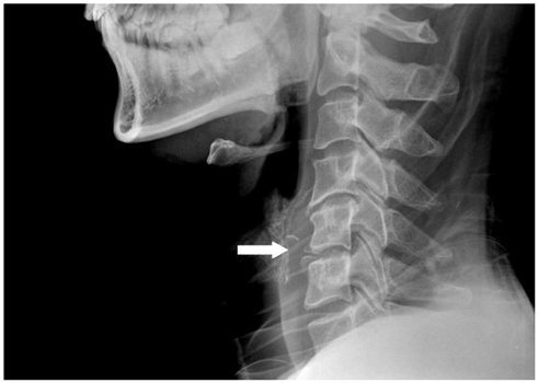
Fig. 2 Lateral view of the cervical spine. There is an anterior bony projection between C5 and C6. The bony mass arises posterior to the anterior margins of the vertebral bodies in the region of the transverse process (arrow).

Fig. 3 CT scan of the cervical spine. The bony projections arose from the anterior tubercles of the right transverse processes (arrows). There is no evidence of bone fracture. A. Axial scan at the level of C5. B. Axial scan at the level of C6.
Reference
-
1. Lapayowker MS. An unusual variant of the cervical spine. Am J Roentgenol Radium Ther Nucl Med. 1960; 83:656–659.2. Applbaum Y, Gerard P, Bryk D. Elongation of the anterior tubercle of a cervical vertebral transverse process: an unusual variant. Skeletal Radiol. 1983; 10:265–267.3. Grilliot JR, Wiles MJ. Elongation of the anterior tubercle of a cervical vertebral transverse process. J Manipulative Physiol Ther. 1988; 11:221–223.4. Mellado JM, Larrosa R, Martín J, Yanguas N, Solanas S, Cozcolluela MR. MDCT of variations and anomalies of the neural arch and its processes: part 1--pedicles, pars interarticularis, laminae, and spinous process. AJR Am J Roentgenol. 2011; 197:W104–W113.5. Mellado JM, Larrosa R, Martín J, Yanguas N, Solanas S, Cozcolluela MR. MDCT of variations and anomalies of the neural arch and its processes: part 2--articular processes, transverse processes, and high cervical spine. AJR Am J Roentgenol. 2011; 197:W114–W121.6. Coley BD. Caffey's Pediatric Diagnostic Imaging. 12th ed. Philadelphia, PA: Mosby/Elsevier;2013. p. 1356–1427.7. Bron JL, van Royen BJ, Wuisman PI. The clinical significance of lumbosacral transitional anomalies. Acta Orthop Belg. 2007; 73:687–695.
- Full Text Links
-
- Actions
-
Cited
- CITED
-
- Close
- Share
-
- Similar articles
-
- Effectiveness of Doppler Image of the Vertebral Artery as an Anatomical Landmark for Identification of Ultrasound-Guided Target Level in Cervical Spine
- Radiologic Characteristics of Vertebral Artery Injury in the Cervical Spine Fracture
- Aberrant Right Vertebral Artery Originating from the Aortic Arch Distal to the Left Subclavian Artery: A Case Report
- Two Cases of Aberrant Vertebral Artery Originating from Aortic Arch Distal to Left Subclavian Artery
- Variations in Entrance of Vertebral Artery in Korean Cervical Spine: MDCT-based Analysis
