J Korean Soc Transplant.
2015 Sep;29(3):160-165. 10.4285/jkstn.2015.29.3.160.
Treatment of Renal Transplant Recipients with Concurrent Acute Cellular Rejection and Transplant Renal Artery Stenosis
- Affiliations
-
- 1Department of Internal Medicine, Bong Seng Memorial Hospital, Busan, Korea. kidney119@hotmail.com
- 2Department of Pathology, Yeungnam University College of Medicine, Daegu, Korea.
- KMID: 2202588
- DOI: http://doi.org/10.4285/jkstn.2015.29.3.160
Abstract
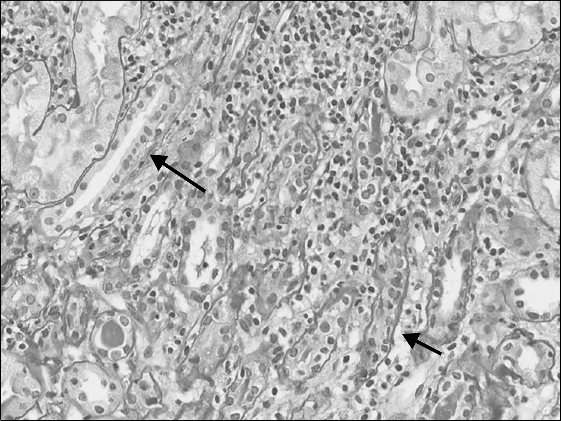
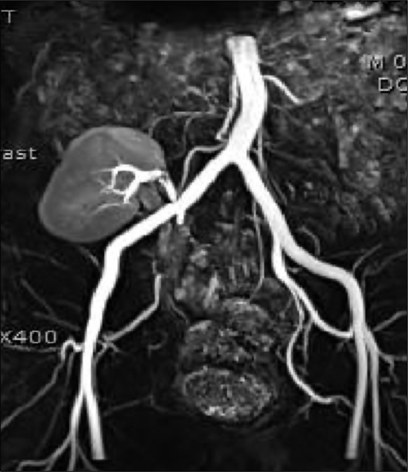
- Transplant renal artery stenosis (TRAS) is a common surgical complication after kidney transplantation (KTP) and is the cause of allograft dysfunction. TRAS is a potentially curable cause of refractory hypertension and allograft dysfunction which accounts for approximately 1% to 5% of cases of post-transplant hypertension. Acute cellular rejection (ACR) is also common after KTP, which is the main cause of allograft dysfunction. Although the incidence of ACR has declined with the advent of new immunosuppressive drugs, it is still around 15% worldwide. Although each disease is frequently seen individually, seeing both together is rare. A 42-year-old man with end stage renal disease underwent KTP, and the donor was his younger brother. Four months after KTP, his serum creatinine was increased to 2.1 mg/dL, and renal biopsy showed interstitial lymphocytic infiltration and tubulitis. With the diagnosis of acute T-cell mediated rejection, steroid pulsing therapy was started, but it was resisted. Therefore thymoglobulin 60 mg (1 mg/kg/day) was administered for 6 days, but serum creatinine was 1.8 mg/dL. Abdomen magnetic resonance angiography showed TRAS, stenosis at the anastomosis site and lobar artery in the lower pole. Percutaneous transluminal angiography was performed successfully. After balloon angioplasty, the stenotic lesion showed a normal size and blood flow. The patient's renal function returned to normal levels and he is currently being followed up for 9 months.
MeSH Terms
-
Abdomen
Adult
Allografts
Angiography
Angioplasty, Balloon
Arteries
Biopsy
Constriction, Pathologic
Creatinine
Diagnosis
Humans
Hypertension
Incidence
Kidney Failure, Chronic
Kidney Transplantation
Magnetic Resonance Angiography
Renal Artery Obstruction*
Renal Artery*
Siblings
T-Lymphocytes
Tissue Donors
Transplantation*
Creatinine
Figure
Reference
-
References
1). Troppmann C, Gillingham KJ, Benedetti E, Almond PS, Gruessner RW, Najarian JS, et al. Delayed graft function, acute rejection, and outcome after cadaver renal transplantation. The multivariate analysis. Transplantation. 1995; 59:962–8.2). Nankivell BJ, Allen RD, O'Connell PJ, Chapman JR. Renal dysfunction in acute rejection. Effect of HLA typing, therapy, and histology. Transplantation. 1995; 60:28–36.3). Djamali A, Samaniego M, Muth B, Muehrer R, Hofmann RM, Pirsch J, et al. Medical care of kidney transplant recipients after the first posttransplant year. Clin J Am Soc Nephrol. 2006; 1:623–40.
Article4). Bruno S, Remuzzi G, Ruggenenti P. Transplant renal artery stenosis. J Am Soc Nephrol. 2004; 15:134–41.5). Roberts JP, Ascher NL, Fryd DS, Hunter DW, Dunn DL, Payne WD, et al. Transplant renal artery stenosis. Transplantation. 1989; 48:580–3.6). Polak WG, Jezior D, Garcarek J, Chudoba P, Patrzalek D, Boratynska M, et al. Incidence and outcome of transplant renal artery stenosis: single center experience. Transplant Proc. 2006; 38:131–2.
Article7). Racusen LC, Solez K, Colvin RB, Bonsib SM, Castro MC, Cavallo T, et al. The Banff 97 working classification of renal allograft pathology. Kidney Int. 1999; 55:713–23.
Article8). Solez K, Axelsen RA, Benediktsson H, Burdick JF, Cohen AH, Colvin RB, et al. International standardization of criteria for the histologic diagnosis of renal allograft rejection: the Banff working classification of kidney transplant pathology. Kidney Int. 1993; 44:411–22.
Article9). Ganji MR, Broumand B. Acute cellular rejection. Iran J Kidney Dis. 2007; 1:54–6.10). Nair MP, Nampoory MR, Said T, Halim MA, Mansour M, Johny KV, et al. Early acute rejection episodes in renal transplantation in relation to immunosuppression protocols: an audit of 100 cases. Transplant Proc. 2005; 37:3029–30.11). Pallardo Mateu LM, Sancho Calabuig A, Capdevila Plaza L, Franco Esteve A. Acute rejection and late renal transplant failure: risk factors and prognosis. Nephrol Dial Transplant. 2004; 19(Suppl 3):iii38–42.
Article12). Madden RL, Mulhern JG, Benedetto BJ, O'Shea MH, Germain MJ, Braden GL, et al. Completely reversed acute rejection is not a significant risk factor for the development of chronic rejection in renal allograft recipients. Transpl Int. 2000; 13:344–50.
Article13). Pascual M, Theruvath T, Kawai T, Tolkoff-Rubin N, Cosimi AB. Strategies to improve longterm outcomes after renal transplantation. N Engl J Med. 2002; 346:580–90.
Article14). Sutherland RS, Spees EK, Jones JW, Fink DW. Renal artery stenosis after renal transplantation: the impact of the hypo-gastric artery anastomosis. J Urol. 1993; 149:980–5.
Article15). Tilney NL, Rocha A, Strom TB, Kirkman RL. Renal artery stenosis in transplant patients. Ann Surg. 1984; 199:454–60.
Article16). Wong W, Fynn SP, Higgins RM, Walters H, Evans S, Deane C, et al. Transplant renal artery stenosis in 77 patients: does it have an immunological cause? Transplantation. 1996; 61:215–9.17). Erley CM, Duda SH, Wakat JP, Sokler M, Reuland P, Muller-Schauenburg W, et al. Noninvasive procedures for diagnosis of renovascular hypertension in renal transplant recipients: a prospective analysis. Transplantation. 1992; 54:863–7.18). Granata A, Clementi S, Londrino F, Romano G, Veroux M, Fiorini F, et al. Renal transplant vascular complications: the role of Doppler ultrasound. J Ultrasound. 2015; 18:101–7.
Article19). Grzelak P, Kurnatowska I, Nowicki M, Muras K, Podgorski M, Strzelczyk J, et al. Detection of transplant renal artery stenosis in the early postoperative period with analysis of parenchymal perfusion with ultrasound contrast agent. Ann Transplant. 2013; 18:187–94.
Article20). Kim SH, An H, Moon SJ, Kim JY, Park SC, Chun HJ, et al. Role of 3D CT-angiography in detecting transplant renal artery stenosis. J Korean Soc Transplant. 2007; 21:88–93.21). Huber A, Heuck A, Scheidler J, Holzknecht N, Baur A, Stangl M, et al. Contrast-enhanced MR angiography in patients after kidney transplantation. Eur Radiol. 2001; 11:2488–95.
Article22). O'Neill W C, Baumgarten DA. Ultrasonography in renal transplantation. Am J Kidney Dis. 2002; 39:663–78.23). Patel NH, Jindal RM, Wilkin T, Rose S, Johnson MS, Shah H, et al. Renal arterial stenosis in renal allografts: retrospective study of predisposing factors and outcome after percutaneous transluminal angioplasty. Radiology. 2001; 219:663–7.
Article24). Leertouwer TC, Gussenhoven EJ, Bosch JL, van Jaarsveld BC, van Dijk LC, Deinum J, et al. Stent placement for renal arterial stenosis: where do we stand? A metaanalysis. Radiology. 2000; 216:78–85.
Article25). Becker GJ, Katzen BT, Dake MD. Noncoronary angioplasty. Radiology. 1989; 170(3 Pt 2):921–40.
Article26). Kuhn FP, Kutkuhn B, Torsello G, Modder U. Renal artery stenosis: preliminary results of treatment with the Strecker stent. Radiology. 1991; 180:367–72.
Article
- Full Text Links
- Actions
-
Cited
- CITED
-
- Close
- Share
- Similar articles
-
- Surgical experience of transplant renal artery stenosis
- Acute Renal Failure in a Renal Allograft Recipient Caused by a Post-Biopsy Renal Arteriovenous Fistula with Transplant Renal Artery Stenosis
- Clinical Significance of Anti-endothelial Cell Antibody in Renal Transplant Recipients
- Clinical significance of copeptin as an early predictor of renal graft dysfunction in renal transplant recipients
- Significance of Resistive index in Renal Transplantation


