J Menopausal Med.
2015 Dec;21(3):155-159. 10.6118/jmm.2015.21.3.155.
Ovarian Cavernous Hemangioma Presenting as a Large Growing Mass in a Postmenopausal Woman: A Case Report and Review of the Literature
- Affiliations
-
- 1Department of Obstetrics and Gynecology, Pusan National University School of Medicine, Busan, Korea. ghkim@pusan.ac.kr
- 2Department of Radiology, Pusan National University School of Medicine, Busan, Korea.
- 3Department of Pathology, Pusan National University School of Medicine, Busan, Korea.
- 4Department of Obstetrics and Gynecology, Kosin University College of Medicine, Busan, Korea.
- KMID: 2200968
- DOI: http://doi.org/10.6118/jmm.2015.21.3.155
Abstract
- Ovarian hemangiomas are usually of the cavernous type, and are rarely encountered. A 73-year-old woman presented with lower abdominal discomfort. Subsequent physical examination depicted a palpable mass in the lower abdomen. Abdominopelvic computed tomography (CT) revealed a well-circumscribed mass with thin septa measuring 12.1 x 9.0 cm in the right ovary. Levels of the tumor markers cancer antigen (CA)-125 and CA 19-9 were within the normal range. At laparoscopy, the tumor was found to be confined to the right ovary and to have a smooth surface. The final histopathological result was ovarian cavernous hemangioma. Microscopically, the mass consisted of multiple, dilated, blood-filled vascular channels separated by loose connective tissue, and all were lined by a single layer of flattened endothelium. The authors present a case of ovarian cavernous hemangioma presenting as a large growing mass in a postmenopausal woman and review previously published literature.
Keyword
MeSH Terms
Figure
-
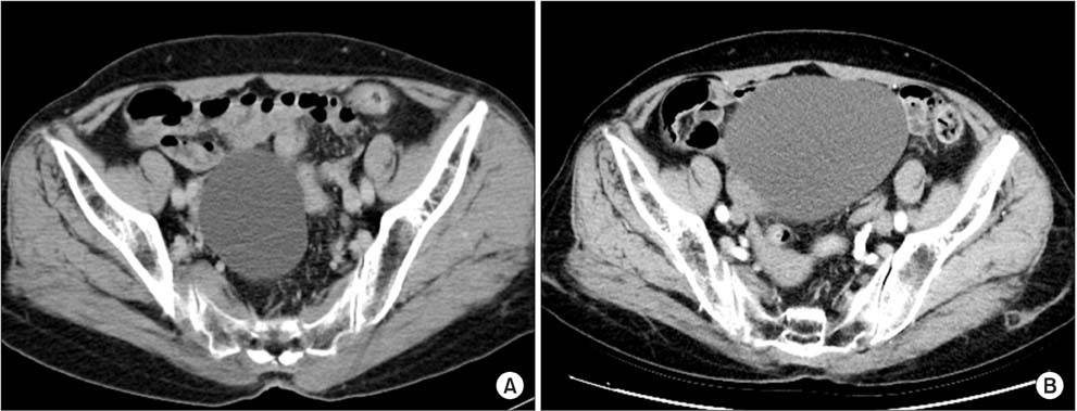
Fig. 1 (A) Contrast enhanced computed tomography (CT) image showing a 6.4 cm, well-defined, thin-walled cyst with thin septa in the right ovary. (B) Follow-up CT image obtained 5 years later showing an increase in mass size to a maximal diameter of 12.1 cm.
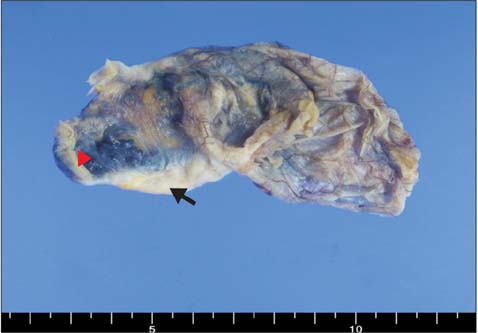
Fig. 2 Gross findings of the ovarian mass. The multiloculated cystic lesion contained occasional blood clots (▸). The remnant ovary is also seen in left lower part (arrow).
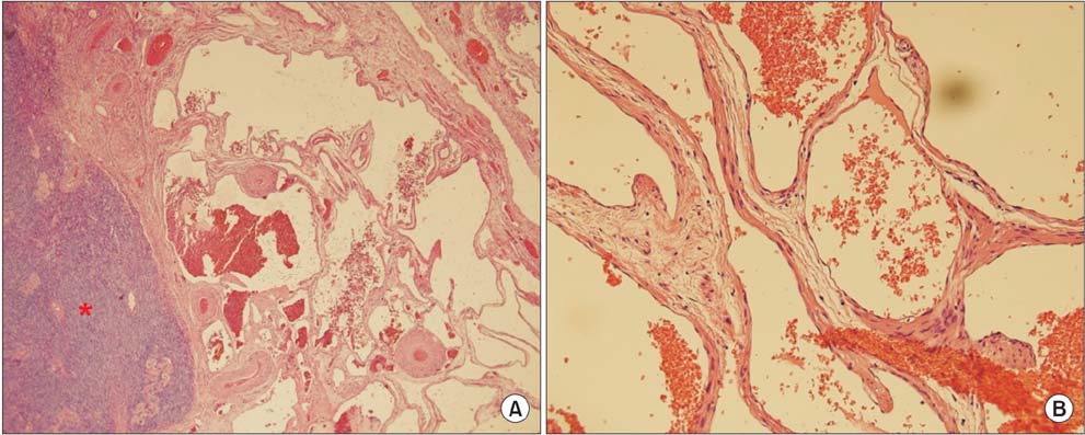
Fig. 3 Microscopic findings of ovarian hemangioma. (A) The mass possessed numerous, dilated vascular spaces adjacent to remnant ovarian tissue on the left side (*) (hematoxylin & eosin [H & E] ×40). (B) Vascular spaces were lined by flattened endothelial cells without cytologic atypia (H & E ×200).
Reference
-
1. Lawhead RA, Copeland LJ, Edwards CL. Bilateral ovarian hemangiomas associated with diffuse abdominopelvic hemangiomatosis. Obstet Gynecol. 1985; 65:597–599.2. Kim MY, Rha SE, Oh SN, Lee YJ, Jung ES, Byun JY. Case report: Ovarian cavernous haemangioma presenting as a heavily calcified adnexal mass. Br J Radiol. 2008; 81:e269–e271.3. Erdemoglu E, Kamaci M, Ozen S, Sahin HG, Kolusari A. Ovarian hemangioma with elevated CA125 and ascites mimicking ovarian cancer. Eur J Gynaecol Oncol. 2006; 27:195–196.4. Kaneta Y, Nishino R, Asaoka K, Toyoshima K, Ito K, Kitai H. Ovarian hemangioma presenting as pseudo-Meigs' syndrome with elevated CA125. J Obstet Gynaecol Res. 2003; 29:132–135.5. Gücer F, Ozyilmaz F, Balkanli-Kaplan P, Mlayim N, Aydin O. Ovarian hemangioma presenting with hyperandrogenism and endometrial cancer: a case report. Gynecol Oncol. 2004; 94:821–824.6. Akbulut M, Bir F, Colakoğlu N, Soysal ME, Düzcan SE. Ovarian hemangioma occurring synchronously with serous papillary carcinoma of the ovary and benign endometrial polyp. Ann Saudi Med. 2008; 28:128–131.7. Carder PJ, Gouldesbrough DR. Ovarian haemangiomas and stromal luteinization. Histopathology. 1995; 26:585–586.8. Uppal S, Heller DS, Majmudar B. Ovarian hemangioma--report of three cases and review of the literature. Arch Gynecol Obstet. 2004; 270:1–5.9. Prat J. Pathology of the ovary. Philadelphia, PA: Saunders;2004.10. Itoh H, Wada T, Michikata K, Sato Y, Seguchi T, Akiyama Y, et al. Ovarian teratoma showing a predominant hemangiomatous element with stromal luteinization: report of a case and review of the literature. Pathol Int. 2004; 54:279–283.11. Talerman A. Nonspecific tumors of the ovary, including mesenchymal tumors and malignant lymphoma. In : Kurman RJ, editor. Blaustein's pathology of the female genital tract. 5th ed. New York, NY: Springer;1987. p. 1035–1062.12. Lee Y. Calcified cavernous hemangioma of the ovary: a case report. J Korean Radiol Soc. 2000; 43:751–754.13. Park CH. Cavernous hemangioma of the ovary. Korean J Obstet Gynecol. 2000; 43:1302–1307.14. Gehrig PA, Fowler WC Jr, Lininger RA. Ovarian capillary hemangioma presenting as an adnexal mass with massive ascites and elevated CA-125. Gynecol Oncol. 2000; 76:130–132.15. Yamawaki T, Hirai Y, Takeshima N, Hasumi K. Ovarian hemangioma associated with concomitant stromal luteinization and ascites. Gynecol Oncol. 1996; 61:438–441.16. Shitsukawa K, Shinomiya S, Tanaka Y, Okada M, Kamada M. Ovarian haemangioma with hyperandrogenism: a case report. J Obstet Gynaecol. 2015; 35:214–215.
