J Lung Cancer.
2009 Dec;8(2):118-119. 10.6058/jlc.2009.8.2.118.
Pulmonary Paragonimiasis Mimicking Lung Malignancy
- Affiliations
-
- 1Lung and Esophageal Cancer Clinic, Chonnam National University Hwasun Hospital, Hawsun, Korea. droij@chonnam.ac.kr
- 2Department of Pulmonary Medicine, Chonnam National University Medical School, Gwangju, Korea.
- 3Department of Pathology, Chonnam National University Medical School, Gwangju, Korea.
- KMID: 2199973
- DOI: http://doi.org/10.6058/jlc.2009.8.2.118
Abstract
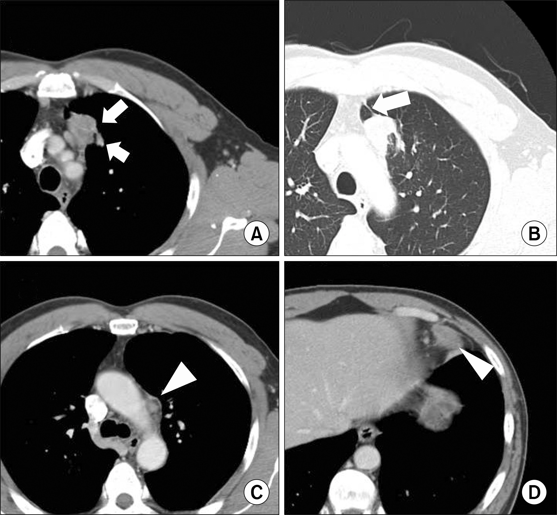
- A 50-year-old man was admitted to our hospital with a complaint of blood-tinged sputum. Chest computed tomography (CT) showed a 37 mm sized heterogeneously lobulated enhancing mass in the central aspect of the left upper lobe and this mass was abutting the adjacent mediastinum (Fig. 1). There was either a focal fibrotic pleural thickening or fissural thickening adjacent to the pulmonary mass. Conglomerated enlarged lymph nodes were observed in the left paraaortic and left anterior cardiophrenic angle areas. 18-Fluorodeoxyglucose positron emission tomography demonstrated hypermetabolic activity in the left upper lobe and the right thyroid gland (Fig. 2). The pathology of the fine needle aspiration from the thyroid gland revealed papillary carcinoma. Since primary or metastatic lung cancer could not be ruled out, video-assisted thoracic surgery was performed. The microscopic findings demonstrated numerous eosinophil infiltrations and many eggs of paragonimiasis westermani, which were observed in the necrotized granuloma area (Fig. 3). He was underwent subtotal thyroidectomy. Moreover, he subsequently received iodine ablation therapy due to papillary carcinoma with regional lymph node invasion.
Keyword
MeSH Terms
Figure
- Full Text Links
- Actions
-
Cited
- CITED
-
- Close
- Share
- Similar articles
-
- A Case of Pulmonary Paragonimiasis Mimicking Lung Cancer Diagnosed by EBUS-TBNA
- A Pulmonary Paragonimiasis Case Mimicking Metastatic Pulmonary Tumor
- A Case of Pulmonary Paragonimiasis Mimicking Pulmonary Tuberculosis
- A Case of Pulmonary Paragonimiasis Presented as Solitary Pulmonary Nodule and Suspected as Lung Cancer on (18)F-Fluorodeoxyglucose Positron Emission Tomography
- A Case of Paragonimiasis Suspected Lung Cancer


