J Korean Acad Prosthodont.
2009 Oct;47(4):424-434. 10.4047/jkap.2009.47.4.424.
Fatigue fracture of different dental implant system under cyclic loading
- Affiliations
-
- 1Department of Prosthodontics, College of Dentistry, Dankook University, Korea. cho8511@dku.edu
- KMID: 2195591
- DOI: http://doi.org/10.4047/jkap.2009.47.4.424
Abstract
- STATEMENT OF PROBLEM: Problems such as loosening and fractures of retained screws and fracture of implant fixture have been frequently reported in implant prosthesis.
PURPOSE: Implant has weak mechanical properties against lateral loading compared to vertical occlusal loading, and therefore, stress analysis of implant fixture depending on its material and geometric features is needed.
MATERIAL AND METHODS: Total 28 of external hexed implants were divided into 7 of 4 groups; Group A (3i, FULL OSSEOTITE(R)Implant), Group B (Nobelbiocare, Branemark System(R)Mk III Groovy RP), Group C (Neobiotec, SinusQuick(TM) EB), Group D (Osstem, US-II). The type III gold alloy prostheses were fabricated using adequate UCLA gold abutments. Fixture, abutment screw, and abutment were connected and cross-sectioned vertically. Hardness test was conducted using MXT-alpha. For fatigue fracture test, with MTS 810, the specimens were loaded to the extent of 60 - 600 N until fracture occurred. The fracture pattern of abutment screw and fixture was observed under scanning electron microscope. A comparative study of stress distribution and fracture area of abutment screw and fixture was carried out through finite element analysis.
RESULTS
1. In Vicker's hardness test of abutment screw, the highest value was measured in group A and lowest value was measured in group D. 2. In all implant groups, implant fixture fractures occurred mainly at the 3 - 4th fixture thread valley where tensile stress was concentrated. When the fatigue life was compared, significant difference was found between the group A, B, C and D (P<.05). 3. The fracture patterns of group B and group D showed complex failure type, a fracture behavior including transverse and longitudinal failure patterns in both fixture and abutment screw. In Group A and C, however, the transverse failure of fixture was only observed. 4. The finite element analysis infers that a fatigue crack started at the fixture surface.
CONCLUSION
The maximum tensile stress was found in the implant fixture at the level of cortical bone. The fatigue fracture occurred when the dead space of implant fixture coincides with jig surface where the maximum tensile stress was generated. To increase implant durability, prevention of surrounding bone resorption is important. However, if the bone resorption progresses to the level of dead space, the frequency of implant fracture would increase. Thus, proper management is needed.
MeSH Terms
Figure
-

Fig. 1. Mean fatigue life of each implant.
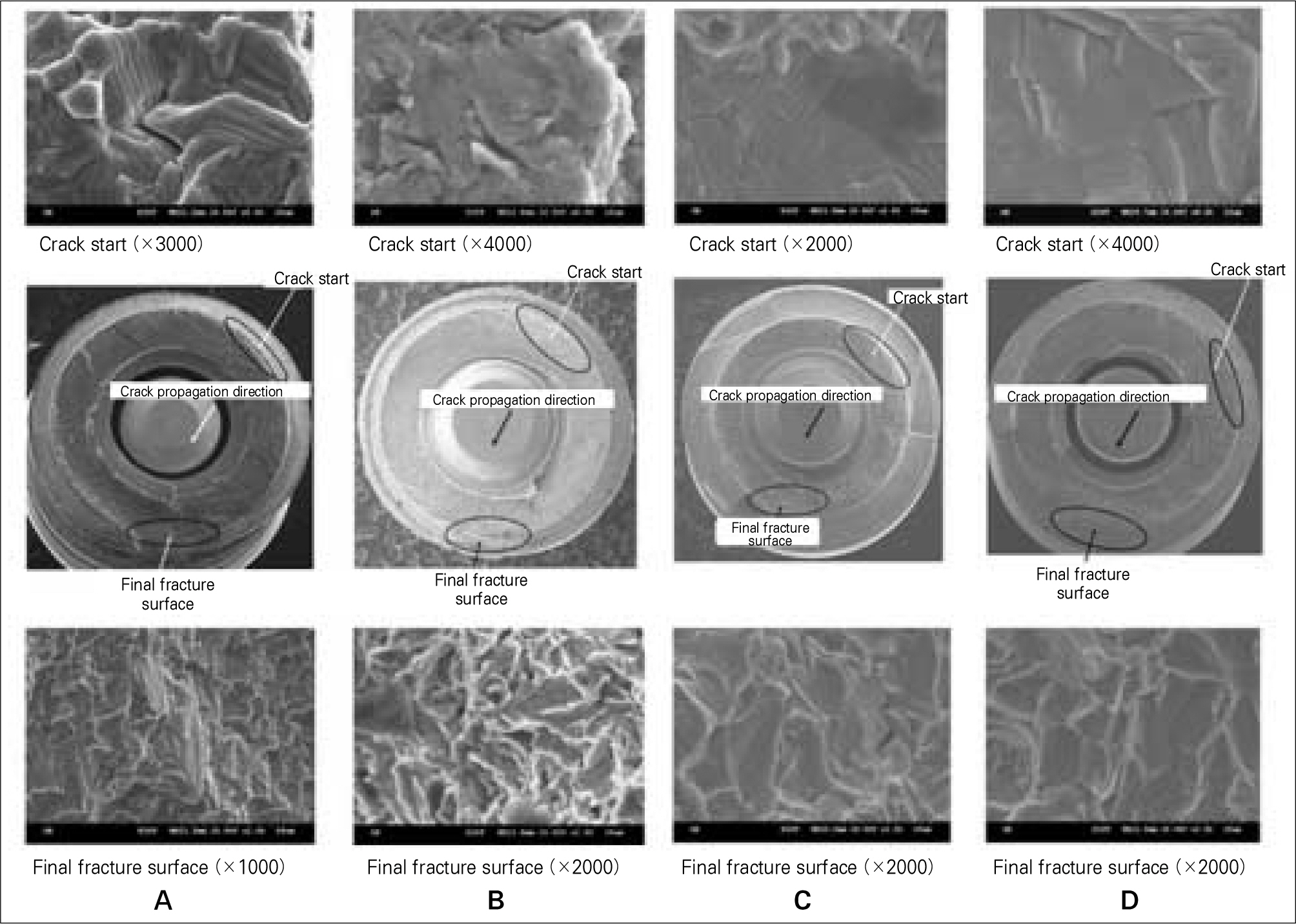
Fig. 2. SEM picture of fractured surface of A, B, C and D implant.
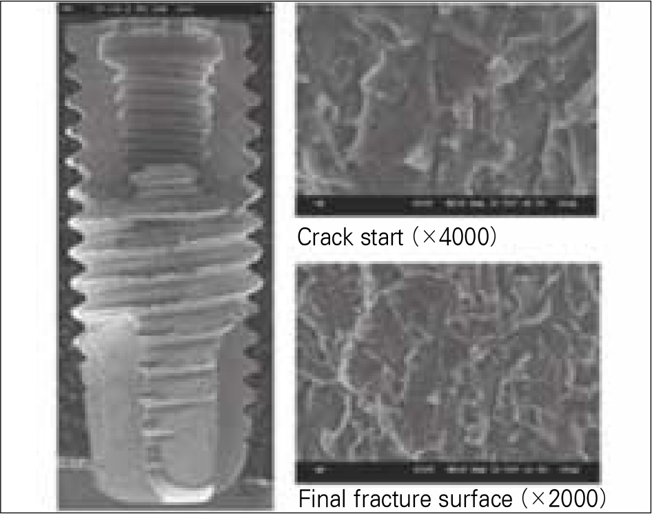
Fig. 3. SEM picture of fractured surface of B implant.
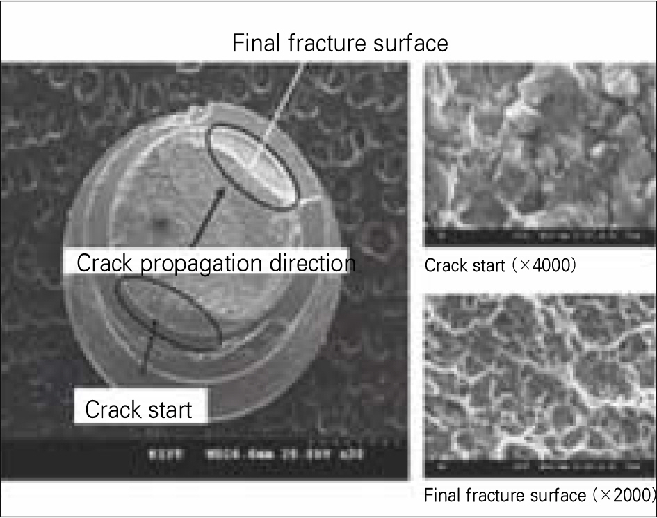
Fig. 4. SEM picture of fracture surface of B abutment screw.
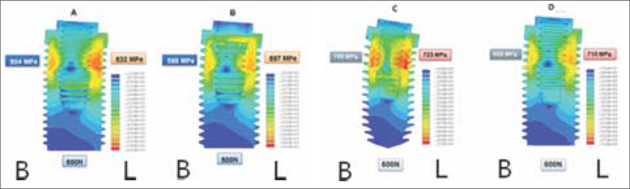
Fig. 5. Stress distribution of implant under 600 N loading in 30° angle.
Cited by 1 articles
-
Comparison of fatigue fracture strength by fixture diameter of mini implants
Yu-Ri Heo, Mee-Kyoung Son, Hee-Jung Kim, Han-Cheol Choe, Chae-Heon Chung
J Korean Acad Prosthodont. 2012;50(3):156-161. doi: 10.4047/jkap.2012.50.3.156.
Reference
-
1.Hoyer SA., Stanford CM., Buranadham S., Fridrich T., Wagner J., Gratton D. Dynamic fatigue properties of the dental implant-abutment interface: joint opening in wide-diameter versus standard-diameter hex-type implants. J Prosthet Dent. 2001. 85:599–607.
Article2.Cibirka RM., Nelson SK., Lang BR., Rueggeberg FA. Examination of the implant-abutment interface after fatigue testing. J Prosthet Dent. 2001. 85:268–75.
Article3.Kim JM., Han JS., Lee SY., Yang JH., Lee JB., Kim YS. A study of screw looswning after dynamic continous fatigue test of several abutment screw. J Korean Acad Prosthodont. 2003. 41:519–31.4.Boggan RS., Strong JT., Misch CE., Bidez MW. Influence of hex geometry and prosthetic table width on static and fatigue strength of dental implants. J Prosthet Dent. 1999. 82:436–40.
Article5.Lim TW., Cho IH., Lim JH., Lim HS. On the fatigue strength of dental implants withdifferent types of connection between fixture and abutment cylinder. J Korean Acad Stomatognathic Func Occlusion. 2002. 18:1–19.6.Adell R., Lekholm U., Rockler B., Bra � nemark PI. A 15-year study of osseointegrated implants in the treatment of the edentulous jaw. Int J Oral Surg. 1981. 10:387–416.
Article7.Balshi TJ. An analysis and management of fractured implants: a clinical report. Int J Oral Maxillofac Implants. 1996. 11:660–6.8.Jemt T., Lekholm U. Oral implant treatment in posterior partially edentulous jaws: a 5-year follow-up report. Int J Oral Maxillofac Implants. 1993. 8:635–40.9.Adell R., Eriksson B., Lekholm U., Bra � nemark PI., Jemt T. Long-term follow-up study of osseointegrated implants in the treatment of totally edentulous jaws. Int J Oral Maxillofac Implants. 1990. 5:347–59.10.Eckert SE., Wollan PC. Retrospective review of 1170 endosseous implants placed in partially edentulous jaws. J Prosthet Dent. 1998. 79:415–21.
Article11.Tagger Green N., Machtei EE., Horwitz J., Peled M. Fracture of dental implants: literature review and report of a case. Implant Dent. 2002. 11:137–43.
Article12.Conrad HJ., Schulte JK., Vallee MC. Fractures related to occlusal overload with single posterior implants: a clinical report. J Prosthet Dent. 2008. 99:251–6.
Article13.Zarb GA., Schmitt A. The longitudinal clinical effectiveness of osseointegrated dental implants: the Toronto study. Part III: Problems and complications encountered. J Prosthet Dent. 1990. 64:185–94.
Article14.Piattelli A., Piattelli M., Scarano A., Montesani L. Light and scanning electron microscopic report of four fractured implants. Int J Oral Maxillofac Implants. 1998. 13:561–4.15.Bonakdarchian M., Askari N., Askari M. Effect of face form on maximal molar bite force with natural dentition. Arch Oral Biol. 2009. 54:201–4.
Article16.Rangert B., Jemt T., Jo ¨rneus L. Forces and moments on Branemark implants. Int J Oral Maxillofac Implants. 1989. 4:241–7.17.Morgan MJ., James DF., Pilliar RM. Fractures of the fixture component of an osseointegrated implant. Int J Oral Maxillofac Implants. 1993. 8:409–14.18.Linkow LI., Donath K., Lemons JE. Retrieval analyses of a blade implant after 231 months of clinical function. Implant Dent. 1992. 1:37–43.
Article19.Siddiqui AA., Claudi R. Proceedings of the 4th International Symposium on Implant Dentistry: Focus on Esthetics. San Diego, California, January 27-29, 1994. Abstracts. J Prosthet Dent. 1994. 72:623–34.20.Gargallo Albiol J., Satorres-Nieto M., Puyuelo Capablo JL., Sa 、nchez Garce 、s MA., Pi Urgell J., Gay Escoda C. Endosseous dental implant fractures: an analysis of 21 cases. Med Oral Patol Oral Cir Bucal. 2008. 13:E124–8.21.Higuchi KW., Folmer T., Kultje C. Implant survival rates in partially edentulous patients: a 3-year prospective multicen-ter study. J Oral Maxillofac Surg. 1995. 53:264–8.22.Parein AM., Eckert SE., Wollan PC., Keller EE. Implant reconstruction in the posterior mandible: a long-term retrospective study. J Prosthet Dent. 1997. 78:34–42.
Article23.Ashley ET., Covington LL., Bishop BG., Breault LG. Ailing and failing endosseous dental implants: a literature review. J Contemp Dent Pract. 2003. 4:35–50.24.Hong WS., Kim TH., Ryu SH., Kook MS., Park HJ., Oh HK. Comparative study of osseoi ntegration of 4 different surfaced implant in the tibia of dogs. J Korean Oral Maxillofac Surg. 2005. 31:46–54.25.Bahat O., Handelsman M. Use of wide implants and double implants in the posterior jaw: a clinical report. Int J Oral Maxillofac Implants. 1996. 11:379–86.26.Han MJ., Chung CH., Choi HC. A study on surface alternation of implant screws after function. J Korean Acad Prosthodont. 2002. 40:275–86.27.Jemt T., Linde 、n B., Lekholm U. Failures and complications in 127 consecutively placed fixed partial prostheses supported by Bra � nemark implants: from prosthetic treatment to first annual checkup. Int J Oral Maxillofac Implants. 1992. 7:40–4.28.Mericske-Stern R., Steinlin Schaffner T., Marti P., Geering AH. Peri-implant mucosal aspects of ITI implants supporting overdentures. A five-year longitudinal study. Clin Oral Implants Res. 1994. 5:9–18.
Article29.Tolman DE., Laney WR. Tissue-integrated prosthesis complications. Int J Oral Maxillofac Implants. 1992. 7:477–84.
Article30.Takeshita F., Suetsugu T., Higuchi Y., Oishi M. Histologic study of failed hollow implants. Int J Oral Maxillofac Implants. 1996. 11:245–50.31.Rangert B., Krogh PH., Langer B., Van Roekel N. Bending overload and implant fracture: a retrospective clinical analysis. Int J Oral Maxillofac Implants. 1995. 10:326–34.32.Carlson B., Carlsson GE. Prosthodontic complications in osseointegrated dental implant treatment. Int J Oral Maxillofac Implants. 1994. 9:90–4.
Article33.Binon PP., McHugh MJ. The effect of eliminating implant/abutment rotational misfit on screw joint stability. Int J Prosthodont. 1996. 9:511–9.34.Santos MD., Pfeifer AB., Silva MR., Sendyk CL., Sendyk WR. Fracture of abutment screw supporting a cemented implant-retained prosthesis with external hexagon connection: a case report with sem evaluation. J Appl Oral Sci. 2007. 15:148–51.
Article35.Vallittu PK. Effect of 180-week water storage on the flexural properties of E-glass and silica fiber acrylic resin composite. Int J Prosthodont. 2000. 13:334–9.36.Kim JH., Lee JB. A comparative study on the correlation between Korean foods and the fractures of PFG and all ceramic crowns for posterior applications. J Korean Acad Prosthodont. 2009. 47:156–63.
Article
- Full Text Links
-
- Actions
-
Cited
- CITED
-
- Close
- Share
-
- Similar articles
-
- The effect of internal implant-abutment connection and diameter on screw loosening
- Pull-off resistance of a screwless implantabutment connection and surface evaluation after cyclic loading
- Influence of tungsten carbide/carbon coating of implant-abutment screw on screw loosening
- The influence of abutment screw tightening timing and DLC coating of conical connection implant system
- Comparison of removal torque between prefabricated and customized abutment screw
