Investigation of osseointegration according to the healing time after having iatrogenic mobility of implant fixtures
- Affiliations
-
- 1Department of Prosthodontics, School of Dentistry, Kyungpook National University, Daegu, Korea. chlee@knu.ac.kr
- KMID: 2195551
- DOI: http://doi.org/10.4047/jkap.2010.48.4.308
Abstract
- PURPOSE
The purpose of this study is to analyze the change in re-osseointegration over time and bone reaction at the interface between implant fixture and the surface of the bone, after destroying re-osseointegration by distorting the bone-implant interface artificially. MATERIAL AND METHODS: Experimental implant fixtures (cp titanium, small ef, Cyrillic3.75 mm x 4 mm) which didn't have surface treatment were produced. Two or three fixtures were implanted on both tibias of twelve female rabbits (New Zealand white, more than 3.5 kg). Then after six weeks, removal torque (RT) was measured and the results were recorded as the first measurement values. The fixtures were submerged again to get reosseointegration between the bone and fixture. To identify the change in re-osseointegration of submerged fixtures over time, six groups had the healing time for four days (group I), one week (group II), two weeks (group III), three weeks (group IV), four weeks (group V) and five weeks (group VI), and then the secondary removal torque was measured for each group. To identify the bone formation under fluorescent light, tetracycline (15 mg/kg, IM) were treated on the rabbits of each group. After the second measurement, the rabbits were sacrificed, and 16 slides were made, two or three for each group. The slides were observed under the fluorescent light with light microscope. To find out the change in the secondary removal torque over the primary removal torque in progress of time, the averages of the increase rate of the primary and secondary torque removal force were calculated. Then, to find out if there were any critical differences between the primary removal torque and the secondary removal torque in each group and among the groups, the results were analyzed statistically by paired t-test, one-way ANOVA, and Duncan's Multiple Range Test.
RESULTS
In group I and II, secondary removal torque decreased, especially in group I. In group III, IV, V, and VI, secondary removal torque increased critically. Comparing the differences among the groups, the critical difference was shown between group I, II and group III, IV, V, VI. Mineralization at the interface between the bone and implant fixture was identified from the first week, and bone formation was shown more clearly from the second week.
CONCLUSION
If the implant fixture remains unforced for a certain period of time after the fixture has had iatrogenic mobility, re-osseointegration occurs at the surface of the fixture, and for tibias of rabbits, higher re-osseointegration was obtained within two weeks.
Keyword
MeSH Terms
Figure
-
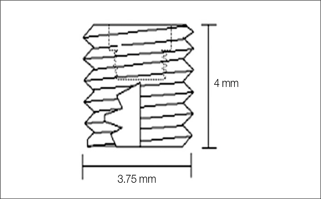
Fig. 1. Schematic diagram of experimental implant.
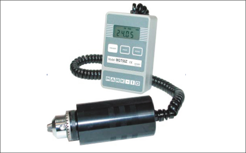
Fig. 2. Digital torque gauge for removal test (Ncm).

Fig. 3. Comparison of 1st and 2nd removal torque values in each group.

Fig. 4. Rate of increase between 1st and 2nd removal torque values in each group.

Fig. 5. Fluorescent finding of each group with light microscope (×20). Arrow indicates the fluorescent labeling.
Cited by 2 articles
-
The influence of intentional mobilization of implant fixtures before osseointegration
Jin-Hyun Cho, Kwang-Heon Jo, Sung-Am Cho, Kyu-Bok Lee, Cheong-Hee Lee
J Korean Acad Prosthodont. 2012;50(3):149-155. doi: 10.4047/jkap.2012.50.3.149.The influence of iatrogenic mobilization in the initial stage of implant installation on final osteointegration
Myeong-Bae Kwak, Jin-Hyun Cho, Du-Heong Lee, Cheong-Hee Lee
J Korean Acad Prosthodont. 2014;52(2):105-112. doi: 10.4047/jkap.2014.52.2.105.
Reference
-
1.Davies JE. Mechanisms of endosseous integration. Int J Prosthodont. 1998. 11:391–401.2.Buser D., Schenk RK., Steinemann S., Fiorellini JP., Fox CH., Stich H. Influence of surface characteristics on bone integration of titanium implants. A histomorphometric study in miniature pigs. J Biomed Mater Res. 1991. 25:889–902.
Article3.Cochran DL., Schenk RK., Lussi A., Higginbottom FL., Buser D. Bone response to unloaded and loaded titanium implants with a sandblasted and acid-etched surface: a histometric study in the canine mandible. J Biomed Mater Res. 1998. 40:1–11.
Article4.Wennerberg A., Albrektsson T., Andersson B. Bone tissue response to commercially pure titanium implants blasted with fine and coarse particles of aluminum oxide. Int J Oral Maxillofac Implants. 1996. 11:38–45.5.Klokkevold PR., Nishimura RD., Adachi M., Caputo A. Osseointegration enhanced by chemical etching of the titanium surface. A torque removal study in the rabbit. Clin Oral Implants Res. 1997. 8:442–7.
Article6.Lazzara RJ., Testori T., Trisi P., Porter SS., Weinstein RL. A human histologic analysis of osseotite and machined surfaces using implants with 2 opposing surfaces. Int J Periodontics Restorative Dent. 1999. 19:117–29.7.Johansson C., Albrektsson T. Integration of screw implants in the rabbit: a 1-year follow-up of removal torque of titanium implants. Int J Oral Maxillofac Implants. 1987. 2:69–75.8.Ivanoff CJ., Sennerby L., Lekholm U. Reintegration of mobilized titanium implants. An experimental study in rabbit tibia. Int J Oral Maxillofac Surg. 1997. 26:310–5.9.Jang JH., Cho JH., Lee CH. Study of the re-osseointegration of implant fixture after mechanical unscrewing. J Korean Acad Prosthodont. 2010. 48:209–14.
Article10.Roberts WE., Turley PK., Brezniak N., Fielder PJ. Implants: Bone physiology and metabolism. CDA J. 1987. 15:54–61.11.Roberts WE. Bone tissue interface. J Dent Educ. 1988. 52:804–9.
Article12.Roberts WE., Smith RK., Zilberman Y., Mozsary PG., Smith RS. Osseous adaptation to continuous loading of rigid endosseous implants. Am J Orthod. 1984. 86:95–111.
Article13.Sennerby L., Thomsen P., Ericson LE. Early tissue response to titanium implants inserted in rabbit cortical bone. J Mater Sci Mater Med. 1993. 4:240–50.
Article
- Full Text Links
-
- Actions
-
Cited
- CITED
-
- Close
- Share
-
- Similar articles
-
- The influence of intentional mobilization of implant fixtures before osseointegration
- Healing of the bone around pure titanium implants without primary bone contact
- Minimal Electrocautery as an Alternative Approach for Implant Removal: A Preliminary Animal Study
- Osseointegration of dental implant in the mandible with diffuse sclerosing osteomyelitis: Report of a rare case
- Influence of cigarette smoke inhalation on dental implant osseointegration in the rat
