The effect of copper alloy scaler tip on the surface roughness of dental implant and restorative materials
- Affiliations
-
- 1Department of Clinical Oral Health Science, Graduate School of Clinical Dentistry, Ewha Womans University, Seoul, Republic of Korea. ekpang@ewha.ac.kr
- 2Department of Periodontology, Mokdong Hospital, Ewha Womans University, Seoul, Republic of Korea.
- 3Department of Periodontology, Graduated School of Medicine, Ewha Womans University, Seoul, Republic of Korea.
- KMID: 2195421
- DOI: http://doi.org/10.4047/jkap.2014.52.3.177
Abstract
- PURPOSE
This study is designed to investigate the various impacts of different types of scaler tips such as cooper alloy base tip and the others on the surface roughness of teeth and implant by the method which is currently in clinical use.
MATERIALS AND METHODS
Four different types of disc shaped porcelain, titanium, zirconia, and Type III gold alloy dental materials sized 15 mm diameter, 1.5 mm thickness were used for the experiment. Plastic hand curette (Group PS), cooper alloy new tip (Group IS), and stainless steel tip (Group SS) were used as testing appliances. A total of 64 specimens were used for this study; Four specimens for each material and appliance group. Surface roughness was formed with 15 degree angle in ultrasonic scaler tip and with 45 degree angle in hand curette of instrument tip and the specimen surface with 5 mm long, one horizontal-reciprocating motion per second for 30 seconds by 40 g force. To survey the surface roughness of each specimen, a field emission scanning electron microscope, an atomic force microscope, and a surface profiler were used. (Ra, microm).
RESULTS
According to SEM, most increased surface roughness was observed in SS group while IS groups had minimal roughness change. Measurement by atomic force microscope presented that the surface roughness of SS group was significantly greater than those of PS, IS and control groups in the type III gold alloy group (P<.05). IS group showed lesser surface roughness changes compared to SS group in porcelain and gold alloy group (P<.05). According to surface profiler, surface roughness of SS group showed greater than those of PS, IS and control groups and IS group showed lesser than those of SS group in all specimen groups. Type III gold alloy group had large changes on surface roughness than those of porcelain, titanium, zirconia (P<.05).
CONCLUSION
The result of this study showed that newly developed copper alloy scaler tip can cause minimal roughness impacts on the surface of implant and dental materials; therefore this may be a useful alternative for prophylaxis of implant and restored teeth.
Keyword
MeSH Terms
Figure
-
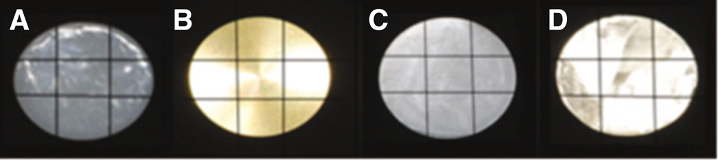
Fig. 1. Experimental specimens of each dental material. (A) Porcelain group, (B) Titanium group, (C) Zirconia group, (D) Gold group.
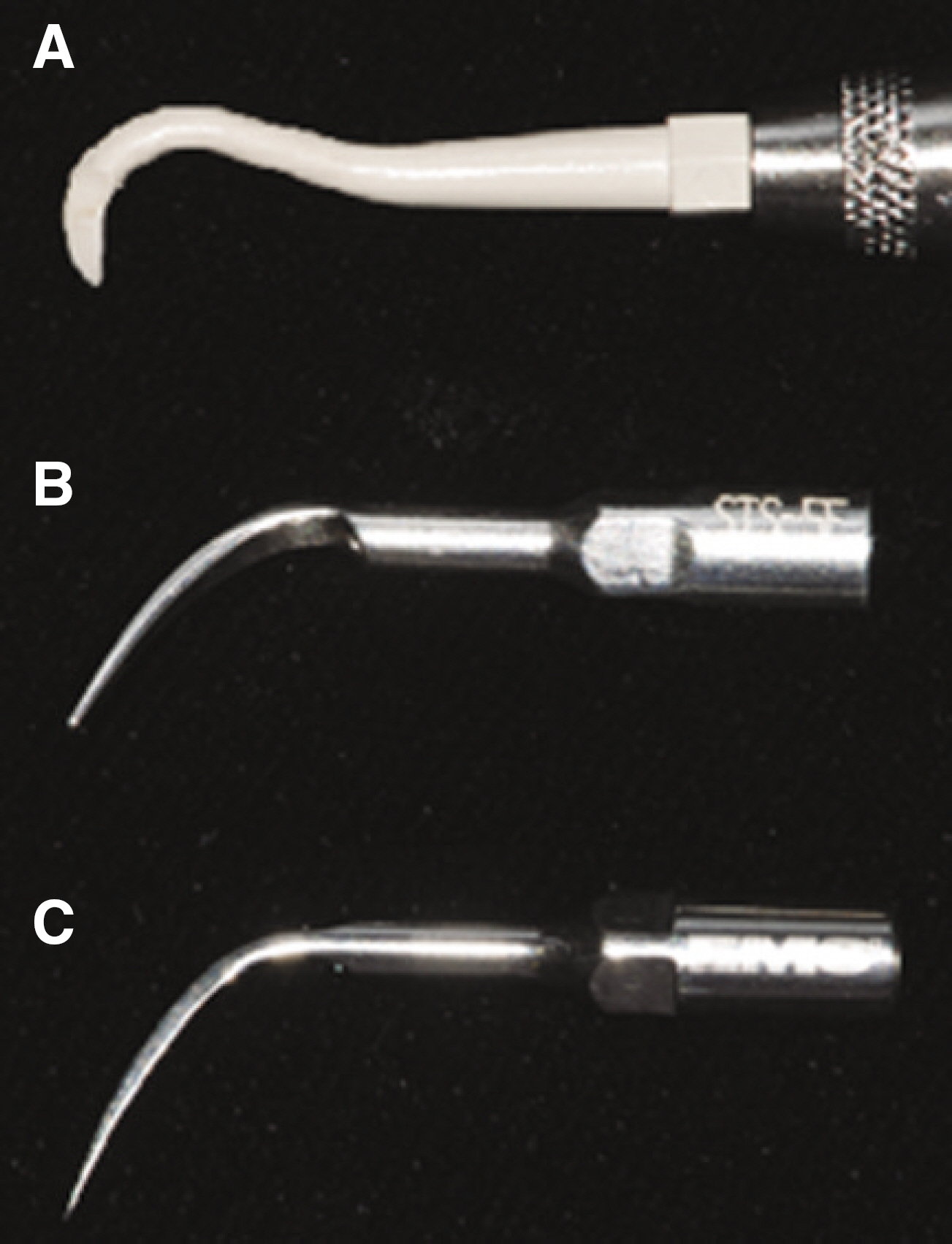
Fig. 2. Scaler tips used in the experiment. (A) Plastic hand curette (PS), (B) Copper alloy scaler tip (IS), (C) Stainless steel scaler tip (SS).
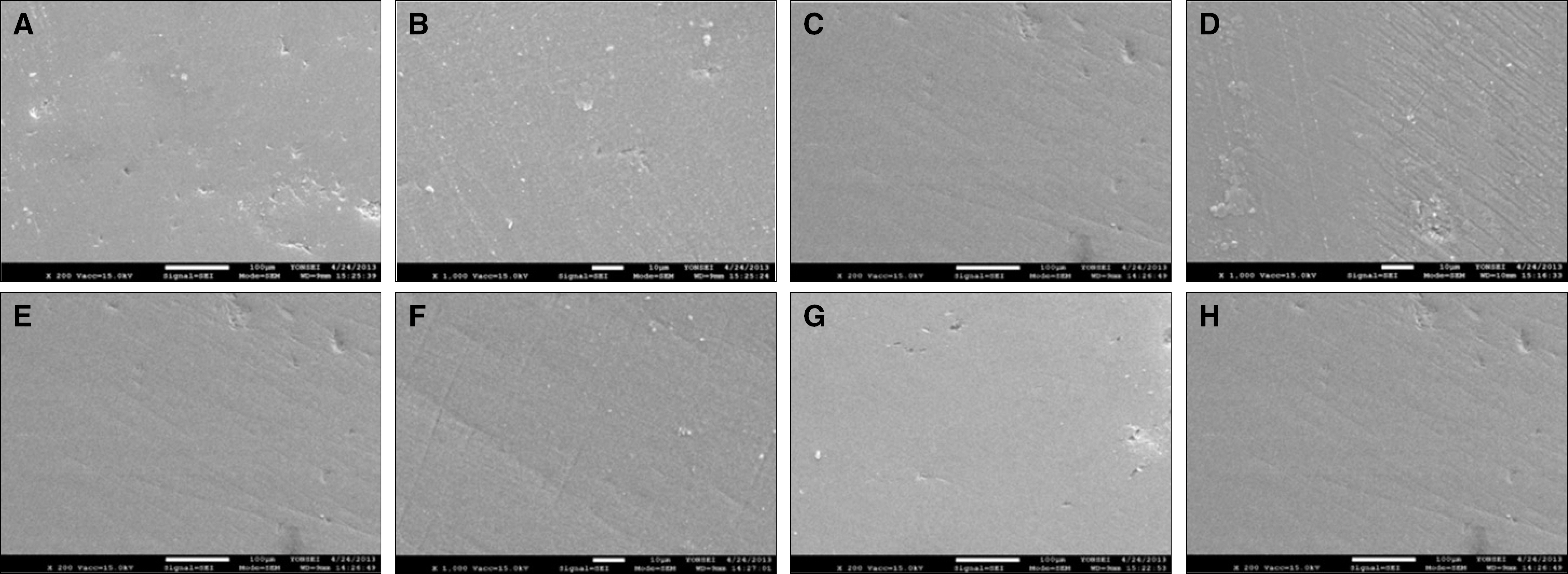
Fig. 3. SEM images of porcelain group. (A),(B) no treatment (Control), (C),(D) Stainless steel scaler tip group (Group SS), (E),(F) Plastic hand curette group (Group PS),(G),(H) Copper alloy scaler tip group (Group IS). Original magnification (A),(C),(E),(G) ×200, (B),(D),(F),(H) ×1000.
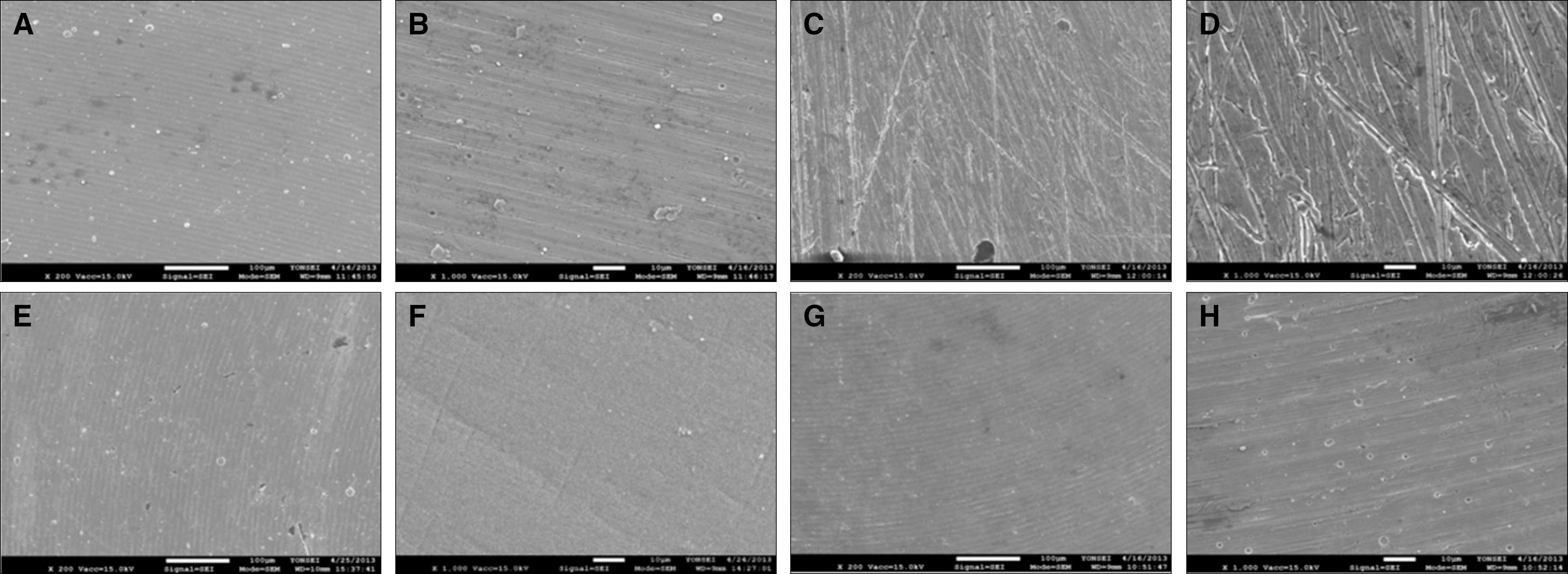
Fig. 4. SEM images of titanium group. (A),(B) no treatment (Control), (C),(D) Stainless steel scaler tip group (Group SS), (E),(F) Plastic hand curette group (Group PS),(G),(H) Copper alloy scaler tip group (Group IS). Original magnification (A),(C),(E),(G) ×200, (B),(D),(F),(H) ×1000.

Fig. 5. SEM images of zirconia group. (A),(B) no treatment (Control), (C),(D) Stainless steel scaler tip group (Group SS), (E),(F) Plastic hand curette group (Group PS),(G),(H) Copper alloy scaler tip group (Group IS). Original magnification (A),(C),(E),(G) ×200, (B),(D),(F),(H) ×1000.

Fig. 6. SEM images of gold group. (A),(B) no treatment (Control), (C),(D) Stainless steel scaler tip group (Group SS), (E),(F) Plastic hand curette group (Group PS), (G),(H) Copper alloy scaler tip group (Group IS). Original magnification (A),(C),(E),(G) ×200, (B),(D),(F),(H) ×1000.
Reference
-
1. Listgarten MA. Pathogenesis of periodontitis. J Clin Periodontol. 1986; 13:418–30.
Article2. Bin AlShaibah WM, El-Shehaby FA, El-Dokky NA, Reda AR. Comparative study on the microbial adhesion to preveneered and stainless steel crowns. J Indian Soc Pedod Prev Dent. 2012; 30:206–11.
Article3. Gristina AG. Biomaterial-centered infection: microbial adhesion versus tissue integration. Science. 1987; 237:1588–95.
Article4. Pameijer CH, Stallard RE, Hiep N. Surface characteristics of teeth following periodontal instrumentation: a scanning electron microscope study. J Periodontol. 1972; 43:628–33.
Article5. Mitchell DF. The irritational qualities of dental materials. J Am Dent Assoc. 1959; 59:954–66.
Article6. Podshadley AG, Harrison JD. Rat connective tissue response to pontic materials. J Prosthet Dent. 1966; 16:110–8.
Article7. Axelsson P, Lindhe J. Effect of controlled oral hygiene procedures on caries and periodontal disease in adults. Results after 6 years. J Clin Periodontol. 1981; 8:239–48.
Article8. Axelsson P, Lindhe J. The significance of maintenance care in the treatment of periodontal disease. J Clin Periodontol. 1981; 8:281–94.
Article9. Renvert S, Roos-Jansa�ker AM, Claffey N. Non-surgical treatment of peri-implant mucositis and peri-implantitis: a literature review. J Clin Periodontol. 2008; 35:305–15.
Article10. Drisko CL, Cochran DL, Blieden T, Bouwsma OJ, Cohen RE, Damoulis P, Fine JB, Greenstein G, Hinrichs J, Somerman MJ, Iacono V, Genco RJ. Research, Science and Therapy Committee of the American Accademy of Periodontology. Position paper: sonic and ultrasonic scalers in periodontics. Research, Science and Therapy Committee of the American Academy of Periodontology. J Periodontol. 2000; 71:1792–801.11. Tunkel J, Heinecke A, Flemmig TF. A systematic review of ef-ficacy of machine-driven and manual subgingival debridement in the treatment of chronic periodontitis. J Clin Periodontol. 2002; 29:72–81.
Article12. Walmsley AD, Lea SC, Landini G, Moses AJ. Advances in pow-er driven pocket/root instrumentation. J Clin Periodontol. 2008; 35:22–8.
Article13. Oda S, Nitta H, Setoguchi T, Izumi Y, Ishikawa I. Current concepts and advances in manual and power-driven instrumentation. Periodontol 2000. 2004; 36:45–58.
Article14. Jang MJ. Evaluation of the neurotoxicity and acute systemic tox-icity of copper ultrasonic scaler tip. Master's Thesis, Department of Prosthodontics, Graduate School, Seoul National University. 2013.15. Kawai K, Urano M. Adherence of plaque components to different restorative materials. Oper Dent. 2001; 26:396–400.16. Kantorski KZ, Scotti R, Valandro LF, Bottino MA, Koga-Ito CY, Jorge AO. Surface roughness and bacterial adherence to resin composites and ceramics. Oral Health Prev Dent. 2009; 7:29–32.17. Thomson-Neal D, Evans GH, Meffert RM. Effects of various prophylactic treatments on titanium, sapphire, and hydroxyap-atite-coated implants: an SEM study. Int J Periodontics Restorative Dent. 1989; 9:300–11.18. Stefani LA. The care and maintenance of the dental implant patient. J Dent Hyg. 1988; 62(447):464–6.19. Rapley JW, Swan RH, Hallmon WW, Mills MP. The surface characteristics produced by various oral hygiene instruments and materials on titanium implant abutments. Int J Oral Maxillofac Implants. 1990; 5:47–52.20. Lee SY, Lai YL, Morgano SM. Effects of ultrasonic scaling and periodontal curettage on surface roughness of porcelain. J Prosthet Dent. 1995; 73:227–32.
Article21. Vigolo P, Motterle M. An in vitro evaluation of zirconia surface roughness caused by different scaling methods. J Prosthet Dent. 2010; 103:283–7.
Article22. Kawashima H, Sato S, Kishida M, Yagi H, Matsumoto K, Ito K. Treatment of titanium dental implants with three piezoelectric ultrasonic scalers: an in vivo study. J Periodontol. 2007; 78:1689–94.
Article23. Hong JW. Effect of wear on tooth surface and efficiency of new ultrasonic scaler tip. Master's Thesis, School of Dentistry, The Graduate School, Seoul National University. 2013.24. Augthun M, Tinschert J, Huber A. In vitro studies on the effect of cleaning methods on different implant surfaces. J Periodontol. 1998; 69:857–64.
Article25. Parham PL Jr, Cobb CM, French AA, Love JW, Drisko CL, Killoy WJ. Effects of an air-powder abrasive system on plasma-sprayed titanium implant surfaces: an in vitro evaluation. J Oral Implantol. 1989; 15:78–86.26. Van de Velde E, Thielens P, Schautteet H, Vanclooster R. Subcutaneous emphysema of the oral floor during cleaning of a bridge fixed on an IMZ implant. Rev Belge Med Dent. 1991; 46:64–71.27. Baek SH, Shon WJ, Bae KS, Kum KY, Lee WC, Park YS. Evaluation of the safety and efficiency of novel metallic ultrasonic scaler tip on titanium surfaces. Clin Oral Implants Res. 2012; 23:1269–74.
Article28. Seol HW, Heo SJ, Koak JY, Kim SK, Baek SH, Lee SY. Surface alterations of several dental materials by a novel ultrasonic scaler tip. Int J Oral Maxillofac Implants. 2012; 27:801–10.29. Gil YM. Effects of ultrasonic scaler tips on the surface of restorative material. Master's Thesis, Department of Prosthodontics, Graduate School, Seoul National University. 2011.30. Kim HC. Effects of various hygiene procedures on the surface characteristics of titanium abutments. Master's Thesis, Esthetic Restorative dentistry, Graduate School of Clinical Dentistry Korea University. 2010.31. Dentkos TR, Berzins DW. Evaluation of cutting efficiency of or-thograde ultrasonic tips by using a nonstatic model. J Endod. 2008; 34:863–5.
Article32. Quirynen M, De Soete M, van Steenberghe D. Infectious risks for oral implants: a review of the literature. Clin Oral Implants Res. 2002; 13:1–19.
Article
- Full Text Links
-
- Actions
-
Cited
- CITED
-
- Close
- Share
-
- Similar articles
-
- Root surface roughness following mechanical instrumentation, in vitro 3 dimensional planimetric study
- Effect of a metallic ultrasonic scaler tip on titanium surfaces: a preliminary study
- Comparative evaluation of roughness of titanium surfaces treated by different hygiene instruments
- The effect of working parameters on removal of casting gold alloy using a piezoelectric ultrasonic scaler with scaler tip in vitro
- A morphologic evaluation of defects created by a piezoelectric ultrasonic scaler on casting gold alloy
