J Korean Soc Surg Hand.
2013 Jun;18(2):59-66. 10.12790/jkssh.2013.18.2.59.
The Short Term Results of All-inside Arthroscopic Repair of the Triangular Fibrocartilage Complex Type 1B Tear by Knotless Suture Anchor
- Affiliations
-
- 1Department of Orthopedic Surgery, Sangmoo Hospital, Gwangju, Korea. scy1art@naver.com
- 2Department of Orthopedic Surgery, Chonnam National University Medical School, Gwangju, Korea.
- KMID: 2194158
- DOI: http://doi.org/10.12790/jkssh.2013.18.2.59
Abstract
- PURPOSE
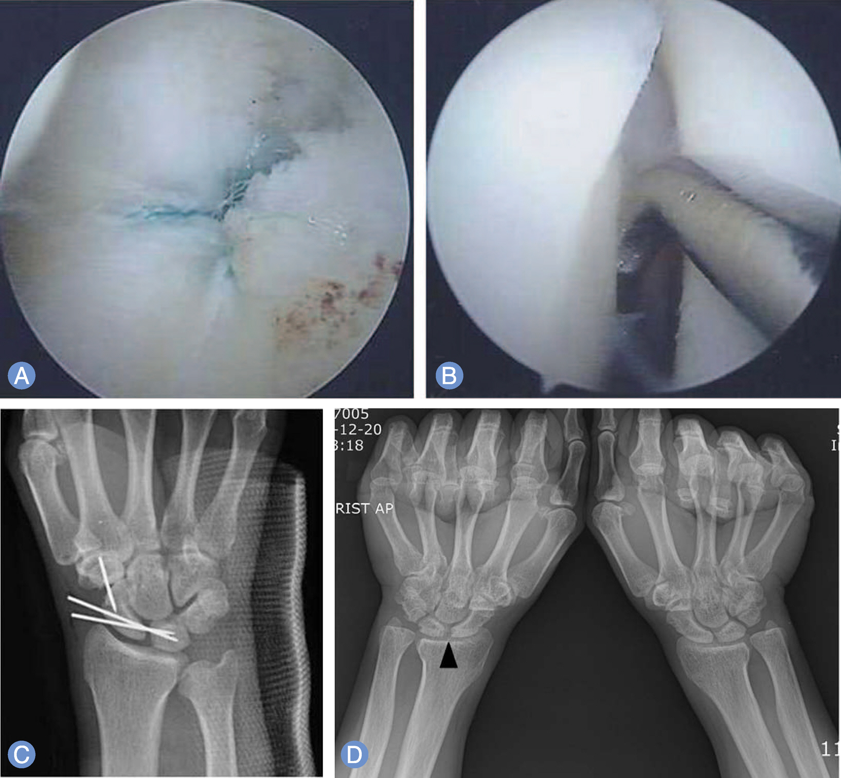
We studied the short term results of the arthroscopic repair of 1B type triangular fibrocartilage complex (TFCC) tear using a knotless suture anchor.
METHODS
We evaluated 23 patients who underwent all-inside arthroscopic TFCC repair using a knotless suture anchor. The average follow-up duration was 6.6 months (range, 3-10 months). Mean duration of symptom was 10.9 months (range, 1 week-7 years). The arthroscopic finding documented 15 type 1B, 2 1B with 1D, and 6 1B with 2C lesions. All showed the positive hook test. The concomitant pathologies were 16 scapholunate injuries and 10 lunotriquetral injuries. TFCC tears were repaired by the knotless suture anchor. The Wafer procedure was done for 2C lesions.
RESULTS
According to Mayo modified wrist score, the result was excellent in 4, good in 14 and fair in 5. Nineteen patients (82.6%) could return to his job or hobby.
CONCLUSION
The all-inside arthroscopic repair using knotless suture anchor for TFCC 1B tear can provide good results. The appropriate management should be done for the concomitant pathologies for the better results.
Figure
Reference
-
1. Palmer AK. Triangular fibrocartilage complex lesions: a classification. J Hand Surg Am. 1989; 14:594–606.
Article2. Corso SJ, Savoie FH, Geissler WB, Whipple TL, Jiminez W, Jenkins N. Arthroscopic repair of peripheral avulsions of the triangular fibrocartilage complex of the wrist: a multicenter study. Arthroscopy. 1997; 13:78–84.
Article3. de Araujo W, Poehling GG, Kuzma GR. New Tuohy needle technique for triangular fibrocartilage complex repair: preliminary studies. Arthroscopy. 1996; 12:699–703.
Article4. Estrella EP, Hung LK, Ho PC, Tse WL. Arthroscopic repair of triangular fibrocartilage complex tears. Arthroscopy. 2007; 23:729–37.
Article5. Nakamura T, Makita A. The proximal ligamentous component of the triangular fibrocartilage complex. J Hand Surg Br. 2000; 25:479–86.
Article6. Nakamura T, Sato K, Okazaki M, Toyama Y, Ikegami H. Repair of foveal detachment of the triangular fibrocartilage complex: open and arthroscopic transosseous techniques. Hand Clin. 2011; 27:281–90.
Article7. Atzei A. New trends in arthroscopic management of type 1-B TFCC injuries with DRUJ instability. J Hand Surg Eur Vol. 2009; 34:582–91.
Article8. Chou KH, Sarris IK, Sotereanos DG. Suture anchor repair of ulnar-sided triangular fibrocartilage complex tears. J Hand Surg Br. 2003; 28:546–50.
Article9. Geissler WB. Arthroscopic knotless peripheral triangular fibrocartilage repair. J Hand Surg Am. 2012; 37:350–5.
Article10. Geissler WB, Freeland AE, Savoie FH, McIntyre LW, Whipple TL. Intracarpal soft-tissue lesions associated with an intra-articular fracture of the distal end of the radius. J Bone Joint Surg Am. 1996; 78:357–65.
Article11. del Pinal F, Studer A, Thams C, Glasberg A. An all-inside technique for arthroscopic suturing of the volar scapholunate ligament. J Hand Surg Am. 2011; 36:2044–6.12. Mathoulin CL, Dauphin N, Wahegaonkar AL. Arthroscopic dorsal capsuloligamentous repair in chronic scapholunate ligament tears. Hand Clin. 2011; 27:563–72.
Article13. Ruch DS, Anderson SR, Ritter MR. Biomechanical comparison of transosseous and capsular repair of peripheral triangular fibrocartilage tears. Arthroscopy. 2003; 19:391–6.
Article14. Anderson ML, Larson AN, Moran SL, Cooney WP, Amrami KK, Berger RA. Clinical comparison of arthroscopic versus open repair of triangular fibrocartilage complex tears. J Hand Surg Am. 2008; 33:675–82.
Article15. Wolf MB, Haas A, Dragu A, et al. Arthroscopic repair of ulnar-sided triangular fibrocartilage complex (Palmer Type 1B) tears: a comparison between short- and midterm results. J Hand Surg Am. 2012; 37:2325–30.
Article16. Ruch DS, Papadonikolakis A. Arthroscopically assisted repair of peripheral triangular fibrocartilage complex tears: factors affecting outcome. Arthroscopy. 2005; 21:1126–30.
Article17. De Smet L, Fabry G. Orientation of the sigmoid notch of the distal radius: determination of different types of the distal radioulnar joint. Acta Orthop Belg. 1993; 59:269–72.18. Papapetropoulos PA, Wartinbee DA, Richard MJ, Leversedge FJ, Ruch DS. Management of peripheral triangular fibrocartilage complex tears in the ulnar positive patient: arthroscopic repair versus ulnar shortening osteotomy. J Hand Surg Am. 2010; 35:1607–13.
Article19. Beredjiklian PK, Bozentka DJ, Leung YL, Monaghan BA. Complications of wrist arthroscopy. J Hand Surg Am. 2004; 29:406–11.
Article20. McAdams TR, Hentz VR. Injury to the dorsal sensory branch of the ulnar nerve in the arthroscopic repair of ulnar-sided triangular fibrocartilage tears using an inside-out technique: a cadaver study. J Hand Surg Am. 2002; 27:840–4.
Article
- Full Text Links
- Actions
-
Cited
- CITED
-
- Close
- Share
- Similar articles
-
- Surgical Technique for Repairing Foveal Tear of the Triangular Fibrocartilage Complex: Arthroscopic Knotless Repair
- Open Repair of Triangular Fibrocartilage Complex Type 1B Tear
- Current Treatment of Triangular Fibrocartilage Complex Injuries
- Surgical Techniques for Repairing Foveal Tear of the Triangular Fibrocartilage Complex: Arthroscopic Transosseous Repair
- Arthroscopic Repair for Traumatic Peripheral Tear of Triangular Fibrocartilage Complex

