Rapidly Enlarging Pediatric Cortical Ependymoma
- Affiliations
-
- 1Department of Neurosurgery, Faculty of Medicine, University of Miyazaki, Miyazaki, Japan. kouji_yamasaki@med.miyazaki-u.ac.jp
- KMID: 2191261
- DOI: http://doi.org/10.3340/jkns.2015.57.6.487
Abstract
- We report a 10-year-old boy with supratentorial cortical ependymoma that rapidly grew in the course of 3 years. He suffered generalized seizures when he was 5 years old; MRI showed a small cortical lesion in the right postcentral gyrus. MRI performed 2 years later revealed no changes. For the next 3 years he was free of seizures. However, at the age of 10 he again suffered generalized seizures and MRI disclosed a large parietal tumor. It was resected totally and he remains free of neurological deficits. The histopathological diagnosis was ependymoma. Pediatric supratentorial cortical ependymomas are extremely rare. We recommend including cortical ependymoma as a differential diagnosis in pediatric patients with cortical mass lesions presenting with seizures and careful follow-up even in the absence of symptoms because these tumors may progress.
MeSH Terms
Figure
-
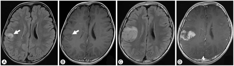
Fig. 1 A and B : MRI scan obtained in 2007 showing a small 14 mm diameter cortical lesion in the right postcentral gyrus. It was hyperintense on the FLAIR image (A), and not gadolinium enhanced (B). C and D : FLAIR image obtained in 2012 showing a large 4 cm diameter parietal tumor (C). It was heterogeneously gadolinium-enhanced (D). FLAIR : fluid-attenuated inversion recovery.

Fig. 2 Intraoperative photograph demonstrating a reddish-gray tumor on the surface of the parietal lobe. It is clearly demarcated from the surrounding brain tissue.
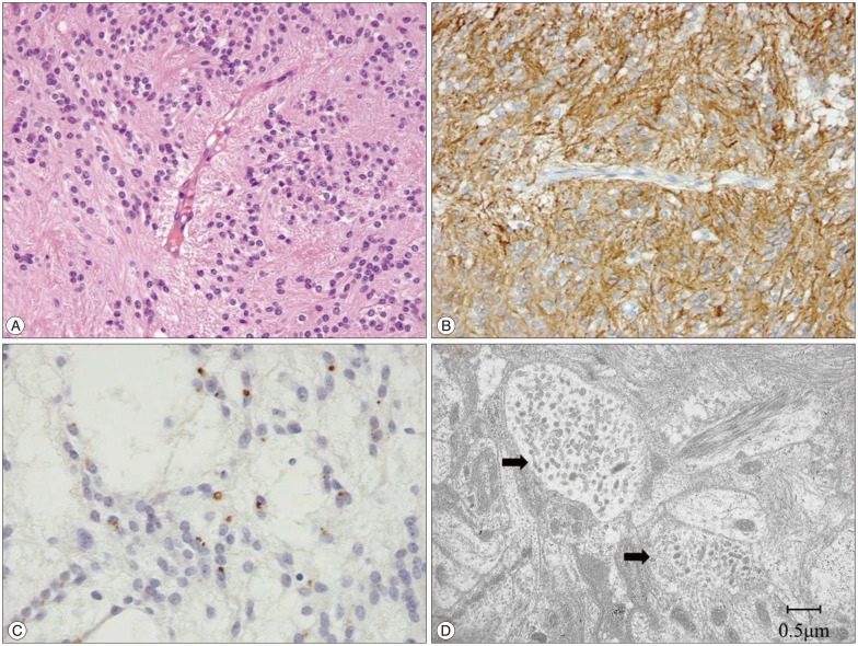
Fig. 3 A : Photomicrographs showing tumor cell proliferation in perivascular pseudorosettes. (HE stain, original magnification ×200). B : Immunohistochemically the tumor cells were positive for glial fibrillary acidic protein (original magnification ×200). C : Ring-like and dot-like epithelial membrane antigen positivity (original magnification ×200). D : Electron micrograph of a tumor cell with a cluster of microvilli (arrow).
Reference
-
1. Afra D, Müller W, Slowik F, Wilcke O, Budka H, Turoczy L. Supratentorial lobar ependymomas : reports on the grading and survival periods in 80 cases, including 46 recurrences. Acta Neurochir (Wien). 1983; 69:243–251. PMID: 6650239.
Article2. Fujimoto K, Ohnishi H, Koshimae N, Ida Y, Kanemoto Y, Motoyama Y, et al. [Brain surface clear cell ependymoma : case report]. No Shinkei Geka. 1999; 27:843–846. PMID: 10478346.3. Ghani AR, Abdullah JM, Ghazali M, Ahmad F, Ahmad KA, Madhavan M. Recurrent paediatric supratentorial extraventricular ependymoma associated with genetic mutation at exon 4 of p53 gene. Singapore Med J. 2008; 49:e192–e194. PMID: 18695856.4. Grajkowska W, Matyja E, Pronicki M, Daszkiewicz P, Roszkowski M, Perek D, et al. Papillary ependymoma with unique superficial cortical location : immunohistochemical and ultrastructural studies. A case report. Folia Neuropathol. 2009; 47:354–361. PMID: 20054788.5. Hamano E, Tsutsumi S, Nonaka Y, Abe Y, Yasumoto Y, Saeki H. Huge supratentorial extraventricular anaplastic ependymoma presenting with massive calcification-case report. Neurol Med Chir (Tokyo). 2010; 50:150–153. PMID: 20185883.
Article6. Hiniker A, Lee HS, Chang S, Berger M, Perry A. Cortical ependymoma with unusual histologic features. Clin Neuropathol. 2013; 32:318–323. PMID: 23458270.
Article7. Lee SK, Lim DJ, Kim SD. Supratentorial cortical ependymoma in a 21-month-old boy. J Korean Neurosurg Soc. 2011; 50:244–247. PMID: 22102957.
Article8. Lehman NL. Patterns of brain infiltration and secondary structure formation in supratentorial ependymal tumors. J Neuropathol Exp Neurol. 2008; 67:900–910. PMID: 18716554.
Article9. Lehman NL, Jorden MA, Huhn SL, Barnes PD, Nelson GB, Fisher PG, et al. Cortical ependymoma. A case report and review. Pediatr Neurosurg. 2003; 39:50–54. PMID: 12784079.10. Lellouch-Tubiana A, Boddaert N, Bourgeois M, Fohlen M, Jouvet A, Delalande O. Angiocentric neuroepithelial tumor (ANET) : a new epilepsy-related clinicopathological entity with distinctive MRI. Brain Pathol. 2005; 15:281–286. PMID: 16389940.
Article11. Liu Z, Li J, Liu Z, Wang Q, Famer P, Mehta A, et al. Supratentorial cortical ependymoma : case series and review of the literature. Neuropathology. 2014; 34:243–252. PMID: 24354554.
Article12. Lum DJ, Halliday W, Watson M, Smith A, Law A. Cortical ependymoma or monomorphous angiocentric glioma? Neuropathology. 2008; 28:81–86. PMID: 18021197.
Article13. Massimino M, Buttarelli FR, Antonelli M, Gandola L, Modena P, Giangaspero F. Intracranial ependymoma : factors affecting outcome. Future Oncol. 2009; 5:207–216. PMID: 19284379.14. Miyazawa T, Hirose T, Nakanishi K, Uozumi Y, Tsuzuki N, Shima K. Supratentorial ectopic cortical ependymoma occurring with intratumoral hemorrhage. Brain Tumor Pathol. 2007; 24:35–40. PMID: 18095143.
Article15. Molina OM, Colina JL, Luzardo GD, Mendez OE, Cardozo D, Velasquez HS, et al. Extraventricular cerebral anaplastic ependymomas. Surg Neurol. 1999; 51:630–635. PMID: 10369231.
Article16. Nakamizo S, Sasayama T, Kondoh T, Inoue S, Shiomi R, Tanaka H, et al. Supratentorial pure cortical ependymoma. J Clin Neurosci. 2012; 19:1453–1455. PMID: 22898199.
Article17. Ohwaki K, Tanishima T, Yoshimasu N, Hojo S, Fujimaki T, Kirino T. [Rapidly enlarging supratentorial ependymoma in a child presenting initially with a small calcified lesion : case report]. No Shinkei Geka. 1997; 25:713–718. PMID: 9266564.18. Prayson RA. Tumours arising in the setting of paediatric chronic epilepsy. Pathology. 2010; 42:426–431. PMID: 20632818.
Article19. Preusser M, Hoischen A, Novak K, Czech T, Prayer D, Hainfellner JA, et al. Angiocentric glioma : report of clinico-pathologic and genetic findings in 8 cases. Am J Surg Pathol. 2007; 31:1709–1718. PMID: 18059228.20. Rigante L, Novello M, Massimi L, Caldarelli M. A cortical cystic epileptogenic lesion : tanycytic ependymoma. Acta Neurol Belg. 2013; 113:523–525. PMID: 23160807.
Article21. Roncaroli F, Consales A, Fioravanti A, Cenacchi G. Supratentorial cortical ependymoma : report of three cases. Neurosurgery. 2005; 57:E192. discussion E192. PMID: 15987557.22. Takeshima H, Kawahara T, Uchida H, Hirano H, Nakazato Y, Kuratsu J. Brain surface ependymoma with repeated episodes of intratumoral hemorrhage-case report. Neurol Med Chir (Tokyo). 2002; 42:166–169. PMID: 12013669.
Article23. Van Gompel JJ, Koeller KK, Meyer FB, Marsh WR, Burger PC, Roncaroli F, et al. Cortical ependymoma : an unusual epileptogenic lesion. J Neurosurg. 2011; 114:1187–1194. PMID: 21235315.24. Vernet O, Farmer JP, Meagher-Villemure K, Montes JL. Supratentorial ectopic ependymoma. Can J Neurol Sci. 1995; 22:316–319. PMID: 8599779.
Article25. Vinchon M, Soto-Ares G, Riffaud L, Ruchoux MM, Dhellemmes P. Supratentorial ependymoma in children. Pediatr Neurosurg. 2001; 34:77–87. PMID: 11287807.
Article26. Wang M, Tihan T, Rojiani AM, Bodhireddy SR, Prayson RA, Iacuone JJ, et al. Monomorphous angiocentric glioma : a distinctive epileptogenic neoplasm with features of infiltrating astrocytoma and ependymoma. J Neuropathol Exp Neurol. 2005; 64:875–881. PMID: 16215459.
Article
