Pure Cortical Ependymoma With RELA-Fusion Positive in a Young Adult
- Affiliations
-
- 1Department of Neurosurgery, Daegu Fatima Hospital, Daegu, Korea
- KMID: 2522229
- DOI: http://doi.org/10.14791/btrt.2021.9.e23
Abstract
- A 25-year-old female presented with a generalized tonic-clonic seizure. She had no previous history of seizures. A brain magnetic resonance imaging scan revealed a solitary enhancing mass in the right fronto-parietal cortex. During surgery, the mass was noted to be pure cortical with no connection to the ventricular lining. The tumor was completely resected. After surgery, the patient had no further seizures. The biopsy result showed a supratentorial ependymoma, which was C11orf95-RELA-fusionpositive.
Figure
-

Fig. 1 Preoperative radiologic finding. A and B: Gadolinium-enhanced T1-weighted brain MRI scans show well enhancing pure cortical mass (white arrows) in size of 3×2×2 cm. C: T2-flair scan shows mass (white arrow) and peri-lesional edema (white arrowhead).
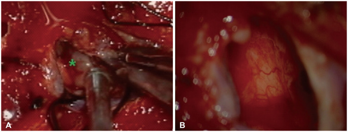
Fig. 2 Intraoperative microscopic finding. A: Intraoperative photograph after dura-mater was opened shows tumor mass (asterisk). The mass was friable solid and highly vascularized, and had no definite adhesion between dura-mater. B: Intraoperative photograph after mass was totally removed shows no definite remnant and parenchymal invasion.

Fig. 3 Histopathological examination with hematoxylin and eosin staining shows a sheet of uniform cells with round nuclei and highly vascularized features (×40) (A) and shows nuclei are with finely dispersed chromatin, clear cytoplasm with well-defined cell membrane, discrete cytoplasmic borders, rare mitosis, no definite necrosis (B, ×100; C, ×200). The black arrows indicate clear cells (C).

Fig. 4 Immunohistochemical examination shows positive with a dot like pattern in EMA staining (×200) (A), focal positive in GFAP (×200) and Olig2 (×200) staining (B and C), and strongly positive in L1CAM staining (×200) (D).
Reference
-
1. Sun S, Wang J, Zhu M, et al. Clinical, radiological, and histological features and treatment outcomes of supratentorial extraventricular ependymoma: 14 cases from a single center. J Neurosurg. 2018; 128:1396–1402. PMID: 28686116.2. Van Gompel JJ, Koeller KK, Meyer FB, et al. Cortical ependymoma: an unusual epileptogenic lesion. J Neurosurg. 2011; 114:1187–1194. PMID: 21235315.3. Sallam YT, Zhang Q, Pandey SK. Cortically based cystic supratentorial RELA fusion-positive ependymoma: a case report with unusual presentation and appearance and review of literature. Radiol Case Rep. 2020; 15:2495–2499. PMID: 33033550.4. Zhu JJ, Jillette N, Li XN, Cheng AW, Lau CC. C11orf95-RELA reprograms 3D epigenome in supratentorial ependymoma. Acta Neuropathol. 2020; 140:951–960. PMID: 32909151.5. Yadav YR, Neha , Chandrakar SK. Pure cortical supratentorial extraventricular ependymoma. Neurol India. 2009; 57:213–215. PMID: 19439861.6. Wu X, Liang P, Zhai X. Supratentorial atypical ectopic ependymoma in a child: a case report. Int J Clin Exp Med. 2017; 10:14827–14833.7. Elsharkawy AE, Abuamona R, Bergmann M, Salem S, Gafumbegete E, Röttger E. Cortical anaplastic ependymoma with significant desmoplasia: a case report and literature review. Case Rep Oncol Med. 2013; 2013:354873. PMID: 24455359.8. Jung TY, Jung S, Kook H, Baek HJ. Treatment decisions of World Health Organization grade II and III ependymomas in molecular era. J Korean Neurosurg Soc. 2018; 61:312–318. PMID: 29742878.9. Pajtler KW, Witt H, Sill M, et al. Molecular classification of ependymal tumors across all CNS compartments, histopathological grades, and age groups. Cancer Cell. 2015; 27:728–743. PMID: 25965575.10. Gupta A, Dwivedi T. A simplified overview of World Health Organization classification update of central nervous system tumors 2016. J Neurosci Rural Pract. 2017; 8:629–641. PMID: 29204027.11. Parker M, Mohankumar KM, Punchihewa C, et al. C11orf95-RELA fusions drive oncogenic NF-κB signalling in ependymoma. Nature. 2014; 506:451–455. PMID: 24553141.12. Pietsch T, Wohlers I, Goschzik T, et al. Supratentorial ependymomas of childhood carry C11orf95-RELA fusions leading to pathological activation of the NF-κB signaling pathway. Acta Neuropathol. 2014; 127:609–611. PMID: 24562983.13. Xia Y, Shen S, Verma IM. NF-κB, an active player in human cancers. Cancer Immunol Res. 2014; 2:823–830. PMID: 25187272.14. Wang Q, Cheng J, Li J, et al. The survival and prognostic factors of supratentorial cortical ependymomas: a retrospective cohort study and literature-based analysis. Front Oncol. 2020; 10:1585. PMID: 32974195.15. Matsumoto Y, Ichikawa T, Kurozumi K, Otani Y, Date I. Clinicopathological and genetic features of supratentorial cortical ependymomas. World Neurosurg. 2019; 129:e417–e428. PMID: 31150846.16. Bijwe S, Ansari S, Jadhav V, Palande D. Pure cortical ependymoma: a rare entity. Asian J Neurosurg. 2015; 10:162–165. PMID: 25972957.17. Berhili S, Aissa A, Kadiri S, et al. Extra-axial ependymoma of the cerebral convexity: a very rare intracranial adult tumor. Neuroradiol J. 2017; 30:281–285. PMID: 28059629.18. Onishi S, Yamasaki F, Nakano Y, et al. RELA fusion-positive anaplastic ependymoma: molecular characterization and advanced MR imaging. Brain Tumor Pathol. 2018; 35:41–45. PMID: 29063976.19. Nakamura T, Fukuoka K, Ikeda J, et al. Encouraging option of multistaged gross total resection for a C11orf-RelA fusion-positive supratentorial anaplastic ependymoma. Brain Tumor Pathol. 2017; 34:160–164. PMID: 28831588.
- Full Text Links
-
- Actions
-
Cited
- CITED
-
- Close
- Share
-
- Similar articles
-
- Aggressive Supratentorial Ependymoma, RELA Fusion-Positive with Extracranial Metastasis: A Case Report
- Rapidly Enlarging Pediatric Cortical Ependymoma
- Supratentorial Cortical Ependymoma in a 21-Month-Old Boy
- Ependymoma Containing Cartilage: A case report
- Acute Paraplegia as a Result of Hemorrhagic Spinal Ependymoma Masked by Spinal Anesthesia: Case Report and Review of Literature
