Quantitative Digital Subtraction Angiography in Pediatric Moyamoya Disease
- Affiliations
-
- 1Department of Radiology, Seoul National University College of Medicine and Institute of Radiation Medicine, Seoul National University Medical Research Center, Seoul, Korea. cheonje@snu.ac.kr
- 2Department of Radiology, Seoul National University Children's Hospital, Seoul, Korea.
- KMID: 2191250
- DOI: http://doi.org/10.3340/jkns.2015.57.6.432
Abstract
- Moyamoya disease is a unique cerebrovascular disorder characterized by idiopathic progressive stenosis at the terminal portion of the internal carotid artery (ICA) and fine vascular network. The aim of this review is to present the clinical application of quantitative digital subtraction angiography (QDSA) in pediatric moyamoya disease. Using conventional angiographic data and postprocessing software, QDSA provides time-contrast intensity curves and then displays the peak time (Tmax) and area under the curve (AUC). These parameters of QDSA can be used as surrogate markers for the hemodynamic evaluation of disease severity and quantification of postoperative neovascularization in moyamoya disease.
Keyword
MeSH Terms
Figure
-
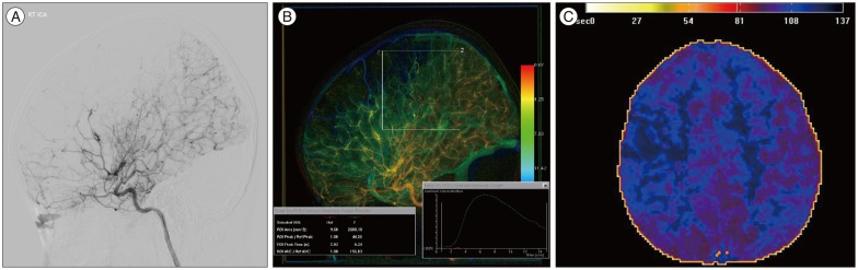
Fig. 1 A 4-year-old boy with left hemiparesis. A : Right internal carotid artery (ICA) angiography shows steno-occlusive change of the distal ICA with basal collateral formation (Suzuki type III). B : Color-coded quantitative DSA (QDSA) demonstrates slow flow (green to blue color) in the right middle cerebral artery (MCA) territory compared to the PCA territory (yellow to orange color). ROI analysis of the MCA territory depicts a time-contrast intensity curve using which Tmax and AUC can be calculated. C : Perfusion MRI of the same patient depicts delayed time to peak in the right MCA territory.
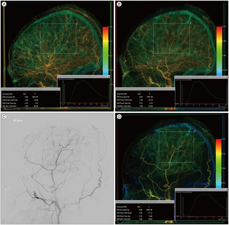
Fig. 2 A 9-year-old girl with left hemiparesis. A and B : Compared to the preoperative quantitative DSA (QDSA) of the right common carotid artery (CCA) (A), postoperative QDSA of the right CCA (B) demonstrates an improved delay of Tmax (7.47 s→5.33 s) and an increased mean AUC. C : Right external carotid artery (ECA) angiography after encephalo-duro-arterio-synangiosis (EDAS) shows good neovascularization (grade 2). D : QDSA of the right ECA angiography after EDAS provides Tmax and AUC of the neovascularization.
Reference
-
1. Hung SC, Liang ML, Lin CF, Lin CJ, Guo WY, Chang FC, et al. New grading of moyamoya disease using color-coded parametric quantitative digital subtraction angiography. J Chin Med Assoc. 2014; 77:437–442. PMID: 25028291.
Article2. Kim SJ, Son TO, Kim KH, Jeon P, Hyun SH, Lee KH, et al. Neovascularization precedes occlusion in moyamoya disease : angiographic findings in 172 pediatric patients. Eur Neurol. 2014; 72:299–305. PMID: 25323466.
Article3. Lin CJ, Hung SC, Guo WY, Chang FC, Luo CB, Beilner J, et al. Monitoring peri-therapeutic cerebral circulation time : a feasibility study using color-coded quantitative DSA in patients with steno-occlusive arterial disease. AJNR Am J Neuroradiol. 2012; 33:1685–1690. PMID: 22499839.
Article4. Matsushima T, Inoue T, Ikezaki K, Matsukado K, Natori Y, Inamura T, et al. Multiple combined indirect procedure for the surgical treatment of children with moyamoya disease. A comparison with single indirect anastomosis and direct anastomosis. Neurosurg Focus. 1998; 5:e4. PMID: 17112207.5. Robertson RL, Burrows PE, Barnes PD, Robson CD, Poussaint TY, Scott RM. Angiographic changes after pial synangiosis in childhood moyamoya disease. AJNR Am J Neuroradiol. 1997; 18:837–845. PMID: 9159360.6. Scott RM, Smith ER. Moyamoya disease and moyamoya syndrome. N Engl J Med. 2009; 360:1226–1237. PMID: 19297575.
Article7. Strother CM, Bender F, Deuerling-Zheng Y, Royalty K, Pulfer KA, Baumgart J, et al. Parametric color coding of digital subtraction angiography. AJNR Am J Neuroradiol. 2010; 31:919–924. PMID: 20167651.
Article8. Suzuki J, Takaku A. Cerebrovascular "moyamoya" disease. Disease showing abnormal net-like vessels in base of brain. Arch Neurol. 1969; 20:288–299. PMID: 5775283.10. Togao O, Mihara F, Yoshiura T, Tanaka A, Noguchi T, Kuwabara Y, et al. Cerebral hemodynamics in Moyamoya disease : correlation between perfusion-weighted MR imaging and cerebral angiography. AJNR Am J Neuroradiol. 2006; 27:391–397. PMID: 16484417.11. Yamada I, Matsushima Y, Suzuki S. Childhood moyamoya disease before and after encephalo-duro-arterio-synangiosis : an angiographic study. Neuroradiology. 1992; 34:318–322. PMID: 1528443.
Article12. Yun TJ, Cheon JE, Na DG, Kim WS, Kim IO, Chang KH, et al. Childhood moyamoya disease : quantitative evaluation of perfusion MR imaging--correlation with clinical outcome after revascularization surgery. Radiology. 2009; 251:216–223. PMID: 19332853.
Article13. Zhang Q, Xu R, Sun Q, Zhang H, Mao J, Shan T, et al. : Exploring the value of using color-coded quantitative DSA evaluation on bilateral common carotid arteries in predicting the reliability of intra-ascending aorta flat detector CT-CBV maps. AJNR Am J Neuroradiol. 2015; [Epub ahead of print].
- Full Text Links
-
- Actions
-
Cited
- CITED
-
- Close
- Share
-
- Similar articles
-
- A Case of Probable Moyamoya Disease (Unilateal Moyamoya Disease) Coexisting Arteriovenous Malformation
- Moyamoya-Like Vasculopathy in Neurosarcoidosis
- Radiologic consideration of intra-arterial digital subtraction angiography
- A Clinical Significance of lntraarterial Digital Subtraction Angiography in Renal Diseases
- Multimodal Imaging Follow-up of a Thrombosed Developmental Venous Anomaly: CT, CT Angiography and Digital Subtraction Angiography
