J Korean Neurosurg Soc.
2015 Feb;57(2):108-113. 10.3340/jkns.2015.57.2.108.
Survival Rates and Risk Factors for Cephalad and L5-S1 Adjacent Segment Degeneration after L5 Floating Lumbar Fusion : A Minimum 2-Year Follow-Up
- Affiliations
-
- 1Department of Neurosurgery, Chung-Ang University College of Medicine, Seoul, Korea. ybkim1218@cau.ac.kr
- KMID: 2191191
- DOI: http://doi.org/10.3340/jkns.2015.57.2.108
Abstract
OBJECTIVE
Although the L5-S1 has distinct structural features in comparison with other lumbar spine segments, not much is known about adjacent segment degeneration (ASD) at the L5-S1 segment. The aim of study was to compare the incidence and character of ASD of the cephalad and L5-S1 segments after L5 floating lumbar fusion.
METHODS
From 2005 to 2010, 115 patients who underwent L5 floating lumber fusion were investigated. The mean follow-up period was 46.1 months. The incidence of radiological and clinical ASD of the cephalad and the L5-S1 segments was compared using survival analysis. Risk factors affecting ASD were analyzed using a log rank test and the Cox proportional hazard model.
RESULTS
Radiological ASD of the L5-S1 segment had a statistically significant higher survival rate than that of the cephalad segment (p=0.001). However, clinical ASD of the L5-S1 segment was significantly lower survival rates than that of the cephalad segment (p=0.038). Risk factor analysis showed that disc degeneration of the cephalad segment and preoperative spinal stenosis of the L5-S1 segment were risk factors.
CONCLUSION
In L5 floating fusion, radiological ASD was more common in the cephalad segment and clinical ASD was more common in the L5-S1 segment. At the L5-S1 segment, the degree of spinal stenosis appears to be the most influential risk factor in ASD incidences, unlike the cephalad segment.
Keyword
MeSH Terms
Figure
-
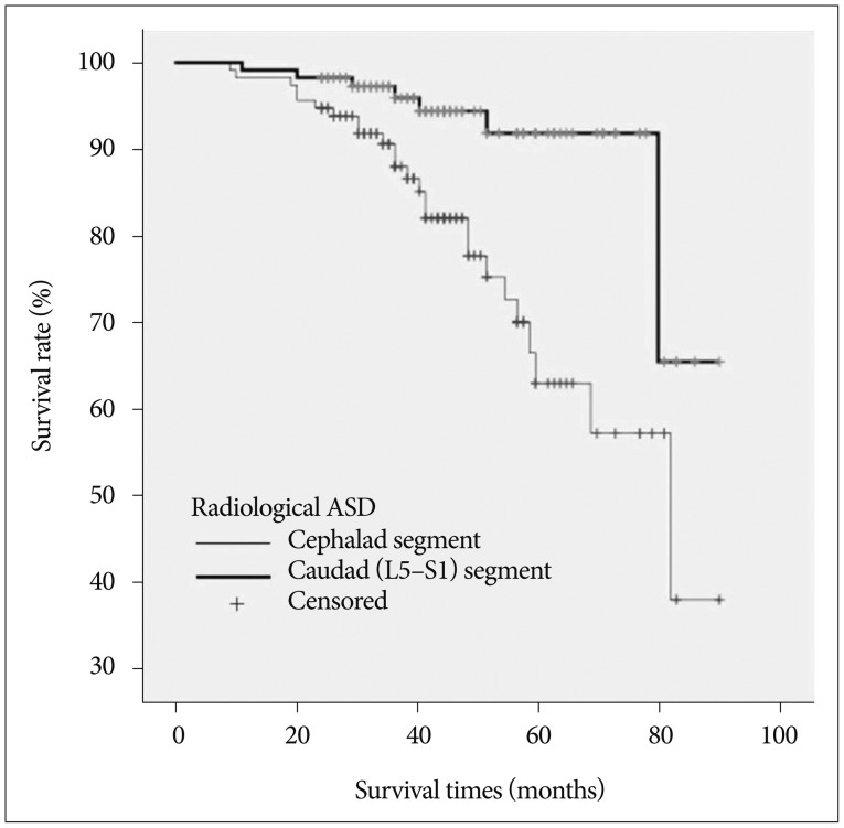
Fig. 1 Kaplan-Meier survivorship curve. The survival rate of the cephalad and caudad (L5-S1) segments for radiological adjacent segment degeneration (ASD) (p=0.001).
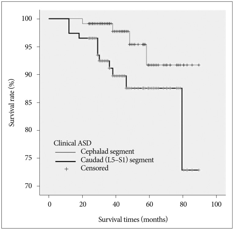
Fig. 2 Kaplan-Meier survivorship curve. The survival rate of the cephalad and caudad (L5-S1) segments for clinical adjacent segment degeneration (ASD) (p=0.038).
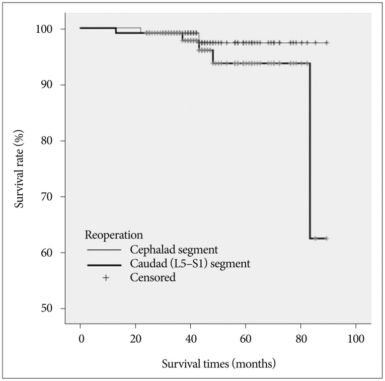
Fig. 3 Kaplan-Meier survivorship curve. The survival rate of the cephalad and caudad (L5-S1) segments for reoperation (p=0.288).
Reference
-
1. Ahn DK, Park HS, Choi DJ, Kim KS, Yang SJ. Survival and prognostic analysis of adjacent segments after spinal fusion. Clin Orthop Surg. 2010; 2:140–147. PMID: 20808584.
Article2. Bridwell KH, Edwards CC 2nd, Lenke LG. The pros and cons to saving the L5-S1 motion segment in a long scoliosis fusion construct. Spine (Phila Pa 1976). 2003; 28:S234–S242. PMID: 14560197.
Article3. Celestre PC, Montgomery SR, Kupperman AI, Aghdasi B, Inoue H, Wang JC. Lumbar clinical adjacent segment pathology: predilection for proximal levels. Spine (Phila Pa 1976). 2014; 39:172–176. PMID: 24153168.4. Cheh G, Bridwell KH, Lenke LG, Buchowski JM, Daubs MD, Kim Y, et al. Adjacent segment disease followinglumbar/thoracolumbar fusion with pedicle screw instrumentation: a minimum 5-year follow-up. Spine (Phila Pa 1976). 2007; 32:2253–2257. PMID: 17873819.
Article5. Ghiselli G, Wang JC, Bhatia NN, Hsu WK, Dawson EG. Adjacent segment degeneration in the lumbar spine. J Bone Joint Surg Am. 2004; 86-A:1497–1503. PMID: 15252099.
Article6. Ghiselli G, Wang JC, Hsu WK, Dawson EG. L5-S1 segment survivorship and clinical outcome analysis after L4-L5 isolated fusion. Spine (Phila Pa 1976). 2003; 28:1275–1280. discussion 1280. PMID: 12811271.
Article7. Imagama S, Kawakami N, Matsubara Y, Kanemura T, Tsuji T, Ohara T. Preventive effect of artificial ligamentous stabilization on the upper adjacent segment impairment following posterior lumbar interbody fusion. Spine (Phila Pa 1976). 2009; 34:2775–2781. PMID: 19940736.
Article8. Kaito T, Hosono N, Mukai Y, Makino T, Fuji T, Yonenobu K. Induction of early degeneration of the adjacent segment after posterior lumbar interbody fusion by excessive distraction of lumbar disc space. J Neurosurg Spine. 2010; 12:671–679. PMID: 20515354.
Article9. Kim TH, Lee BH, Moon SH, Lee SH, Lee HM. Comparison of adjacent segment degeneration after successful posterolateral fusion with unilateral or bilateral pedicle screw instrumentation: a minimum 10-year follow-up. Spine J. 2013; 13:1208–1216. PMID: 24075027.
Article10. Lawrence BD, Wang J, Arnold PM, Hermsmeyer J, Norvell DC, Brodke DS. Predicting the risk of adjacent segment pathology after lumbar fusion: a systematic review. Spine (Phila Pa 1976). 2012; 37(22 Suppl):S123–S132. PMID: 22885827.11. Lee CS, Chung SS, Choi SW, Yu JW, Sohn MS. Critical length of fusion requiring additional fixation to prevent nonunion of the lumbosacral junction. Spine (Phila Pa 1976). 2010; 35:E206–E211. PMID: 20195201.
Article12. Lee CS, Hwang CJ, Lee SW, Ahn YJ, Kim YT, Lee DH, et al. Risk factors for adjacent segment disease after lumbar fusion. Eur Spine J. 2009; 18:1637–1643. PMID: 19533182.
Article13. Leong JC, Luk KD, Chow DH, Woo CW. The biomechanical functions of the iliolumbar ligament in maintaining stability of the lumbosacral junction. Spine (Phila Pa 1976). 1987; 12:669–674. PMID: 3686218.
Article14. Liao JC, Chen WJ, Chen LH, Niu CC, Keorochana G. Surgical outcomes of degenerative spondylolisthesis with L5-S1 disc degeneration: comparison between lumbar floating fusion and lumbosacral fusion at a minimum 5-year follow-up. Spine (Phila Pa 1976). 2011; 36:1600–1677. PMID: 21242863.15. Luk KD, Ho HC, Leong JC. The iliolumbar ligament. A study of its anatomy, development and clinical significance. J Bone Joint Surg Br. 1986; 68:197–200. PMID: 3958002.
Article16. Miyasaka K, Ohmori K, Suzuki K, Inoue H. Radiographic analysis of lumbar motion in relation to lumbosacral stability. Investigation of moderate and maximum motion. Spine (Phila Pa 1976). 2000; 25:732–737. PMID: 10752107.
Article17. Ohmori K, Ishida Y, Takatsu T, Inoue H, Suzuki K. Vertebral slip in lumbar spondylolysis and spondylolisthesis. Long-term follow-up of 22 adult patients. J Bone Joint Surg Br. 1995; 77:771–773. PMID: 7559708.
Article18. Park P, Garton HJ, Gala VC, Hoff JT, McGillicuddy JE. Adjacent segment disease after lumbar or lumbosacral fusion: review of the literature. Spine (Phila Pa 1976). 2004; 29:1938–1944. PMID: 15534420.
Article19. Pearcy M, Portek I, Shepherd J. Three-dimensional x-ray analysis of normal movement in the lumbar spine. Spine (Phila Pa 1976). 1984; 9:294–297. PMID: 6374922.
Article20. Pfirrmann CW, Metzdorf A, Zanetti M, Hodler J, Boos N. Magnetic resonance classification of lumbar intervertebral disc degeneration. Spine (Phila Pa 1976). 2001; 26:1873–1878. PMID: 11568697.
Article21. Sears WR, Sergides IG, Kazemi N, Smith M, White GJ, Osburg B. Incidence and prevalence of surgery at segments adjacent to a previous posterior lumbar arthrodesis. Spine J. 2011; 11:11–20. PMID: 21168094.
Article22. Tan Y, Aghdasi BG, Montgomery SR, Inoue H, Lu C, Wang JC. Kinetic magnetic resonance imaging analysis of lumbar segmental mobility in patients without significant spondylosis. Eur Spine J. 2012; 21:2673–2679. PMID: 22674194.
Article
- Full Text Links
-
- Actions
-
Cited
- CITED
-
- Close
- Share
-
- Similar articles
-
- Incidence and Characteristics of Clinical L5–S1 Adjacent Segment Degeneration after L5 Floating Lumbar Fusion: A Multicenter Study
- Accelerated L5-S1 Segment Degeneration after Spinal Fusion on and above L4-5 : Minimum 4-Year Follow-Up Results
- The Changes of Adjacent Segments after Fusions (above 3-levels to L5) in Degenerative Lumbar Spinal Disorders
- A Comparative Study of the Floating L4-5) vs Lumbosacral L4-S1) Spinal Fusions
- Pseudarthrosis at L5-S1 after Posterolateral Lumbar Fusion
