Primary Malignant Melanoma in the Pineal Region
- Affiliations
-
- 1Department of Neurosurgery, Seoul St. Mary's Hospital, The Catholic University of Korea, Seoul, Korea. jhyun@catholic.ac.kr
- KMID: 2191145
- DOI: http://doi.org/10.3340/jkns.2014.56.6.504
Abstract
- A 59-year-old male patient had 5-month history of gait disturbance and memory impairment. His initial brain computed tomography scan showed 3.5x2.8 cm sized mass with high density in the pineal region. The tumor was hypointense on T2 weighted magnetic resonance images and hyperintense on T1 weighted magnetic resonance images with heterogenous enhancement of central portion. The tumor was totally removed via the occipital transtentorial approach. Black mass was observed in the operation field, and after surgery, histopathological examination confirmed the diagnosis of malignant melanoma. Whole spine magnetic resonance images and whole body 18-fluoro-deoxyglucose positron emission tomography could not demonstrate the primary site of this melanoma. Scrupulous physical examination of his skin and mucosa was done and dark pigmented lesion on his left leg was found, but additional studies including magnetic resonance images and skin biopsy showed negative finding. As a result, final diagnosis of primary pineal malignant melanoma was made. He underwent treatment with the whole brain radiotherapy and extended local boost irradiation without chemotherapy. His preoperative symptoms were disappeared, and no other specific neurological deficits were founded. His follow-up image studies showed no recurrence or distant metastasis until 26 weeks after surgery. Primary pineal malignant melanomas are extremely rare intracranial tumors, and only 17 cases have been reported since 1899. The most recent case report showed favorable outcome by subtotal tumor resection followed by whole brain and extended local irradiation without chemotherapy. Our case is another result to prove that total tumor resection with radiotherapy can be the current optimal treatment for primary malignant melanoma in the pineal region.
MeSH Terms
Figure
-

Fig. 1 Initial computed tomography scan showing a high density mass in the pineal region with minimal obstructive hydrocephalus suspected.
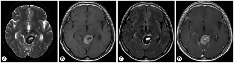
Fig. 2 Preoperative magnetic resonance imaging. The tumor showed dense hypointense rim on T2 weighted images (A) and FLAIR images (C), which was hyperintense on T1 weighted images (B). It contained heterogenous enhancement of central portion (D).

Fig. 3 Intraoperative photograph (left occipital transtentorial approach) showing basal vein of Rosenthal and black tumor mass in the pineal region after opening the tentorium. The tumor was adhered to midbrain.
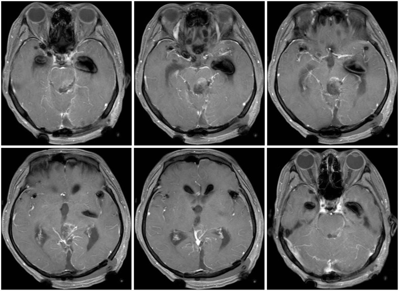
Fig. 4 Postoperative magnetic resonance imaging. Grossly total resection of the tumor was done and there was no evidence of remnant tumor nor ventriculomegaly and no other complications. Only minimal postoperative change was noted at the tumor bed area.

Fig. 5 Histologic finding of tumor specimen. Histopathological examination revealed anaplastic spindled or epithelioid cells with large amounts of melanin pigmentations (H&E ×40).
Reference
-
1. Arantes M, Castro AF, Romão H, Meireles P, Garcia R, Honavar M, et al. Primary pineal malignant melanoma : case report and literature review. Clin Neurol Neurosurg. 2011; 113:59–64. PMID: 20869806.2. Barron J, Morris-Larkin C, Finch T, Maroun F, Hache N, Yousef GM. Long survival of primary pineal melanoma with radiation treatment only. Can J Neurol Sci. 2007; 34:251–253. PMID: 17598609.
Article3. Bookland M, Anderson WS, Biser-Rohrbaugh A, Jallo GI. Primary pineal malignant melanoma. Pediatr Neurosurg. 2007; 43:303–308. PMID: 17627147.
Article4. Bruce JN, Stein BM. Surgical management of pineal region tumors. Acta Neurochir (Wien). 1995; 134:130–135. PMID: 8748771.
Article5. Escott EJ. A variety of appearances of malignant melanoma in the head : a review. Radiographics. 2001; 21:625–639. PMID: 11353111.
Article6. Gaviani P, Mullins ME, Braga TA, Hedley-Whyte ET, Halpern EF, Schaefer PS, et al. Improved detection of metastatic melanoma by T2*-weighted imaging. AJNR Am J Neuroradiol. 2006; 27:605–608. PMID: 16552002.7. Hayward RD. Malignant melanoma and the central nervous system. A guide for classification based on the clinical findings. J Neurol Neurosurg Psychiatry. 1976; 39:526–530. PMID: 950562.
Article8. Isiklar I, Leeds NE, Fuller GN, Kumar AJ. Intracranial metastatic melanoma : correlation between MR imaging characteristics and melanin content. AJR Am J Roentgenol. 1995; 165:1503–1512. PMID: 7484597.
Article9. Landis SH, Murray T, Bolden S, Wingo PA. Cancer statistics, 1998. CA Cancer J Clin. 1998; 48:6–29. PMID: 9449931.
Article10. Martin-Blondel G, Rousseau A, Boch AL, Cacoub P, Sène D. Primary pineal melanoma with leptomeningeal spreading : case report and review of the literature. Clin Neuropathol. 2009; 28:387–394. PMID: 19788056.11. Mitchell PJ, Funt SA, Gonzales MF, Popovic EA. Primary pineal and meningeal malignant melanomatosis. J Clin Neurosci. 1998; 5:353–356. PMID: 18639049.
Article12. Rubino GJ, King WA, Quinn B, Marroquin CE, Verity MA. Primary pineal melanoma : case report. Neurosurgery. 1993; 33:511–515. discussion 515. PMID: 8413885.13. Sampson JH, Carter JH Jr, Friedman AH, Seigler HF. Demographics, prognosis, and therapy in 702 patients with brain metastases from malignant melanoma. J Neurosurg. 1998; 88:11–20. PMID: 9420067.
Article14. Shinsato Y, Hanada T, Kisanuki T, Yonezawa H, Yunoue S, Yoshioka T, et al. Primary malignant melanoma in the pineal region treated without chemotherapy. Surg Neurol Int. 2012; 3:123. PMID: 23226609.
Article15. Suzuki T, Yasumoto Y, Kumami K, Matsumura K, Kumami M, Mochizuki M, et al. Primary pineal melanocytic tumor. Case report. J Neurosurg. 2001; 94:523–527. PMID: 11235961.16. Wang J, Guo ZZ, Wang YJ, Zhang SG, Xing DG. Microsurgery for the treatment of primary malignant intracranial melanoma : a surgical series and literature review. Eur J Surg Oncol. 2014; 40:1062–1071. PMID: 24360613.
Article17. Yamane K, Shima T, Okada Y, Nishida M, Okita S, Hatayama T, et al. Primary pineal melanoma with long-term survival : case report. Surg Neurol. 1994; 42:433–437. PMID: 7974151.
- Full Text Links
-
- Actions
-
Cited
- CITED
-
- Close
- Share
-
- Similar articles
-
- Surgical Treatment of Cavernous Malformation of Pineal Region
- Primary malignant melanoma arising in a cystic teratoma
- A Case of Primary Malignant Melanoma of the Vagina: Trial of a Wide Local Excision of Vagina and Rectum
- Primary Malignant Melanoma of the Cervical Spinal Nerve Root: A Case Report
- Primary malignant melanoma of the esophagus
