Anatomic Feasibility of Posterior Cervical Pedicle Screw Placement in Children : Computerized Tomographic Analysis of Children Under 10 Years Old
- Affiliations
-
- 1Department of Neurosurgery, Incheon St. Mary's Hospital, The Catholic University of Korea, Incheon, Korea.
- 2Department of Neurosurgery, St. Vincent's Hospital, The Catholic University of Korea, Suwon, Korea. jatagi15@paran.com
- KMID: 2191135
- DOI: http://doi.org/10.3340/jkns.2014.56.6.475
Abstract
OBJECTIVE
To evaluate the anatomical feasibility of 3.5 mm screw into the cervical spine in the pediatric population and to establish useful guidelines for their placement.
METHODS
A total of 37 cervical spine computerized tomography scans (24 boys and 13 girls) were included in this study. All patients were younger than 10 years of age at the time of evaluation for the period of 2007-2011.
RESULTS
For the C1 screw placement, entry point height (EPH) was the most restrictive factor (47.3% patients were larger than 3.5 mm). All C2 lamina had a height larger than 3.5 mm and 68.8% (51/74) of C2 lamina had a width thicker than 3.5 mm. For C2 pedicle width, 55.4% (41/74) of cases were larger than 3.5 mm, while 58.1% (43/74) of pedicle heights were larger than 3.5 mm. For pedicle width of subaxial spine, 75.7% (C3), 73% (C4), 82.4% (C5), 89.2% (C6), and 98.1% (C7, 1/54) were greater than 3.5 mm. Mean lamina width of subaxial cervical spine was 3.1 (C3), 2.7 (C4), 2.9 (C5), 3.8 (C6), and 4.0 mm (C7), respectively. Only 34.6% (127/370) of subaxial (C3-7) lamina thickness were greater than 3.5 mm. Mean length of lateral mass for the lateral mass screw placement was 9.28 (C3), 9.08 (C4), 8.81 (C5), 8.98 (C6), and 10.38 mm (C7).
CONCLUSION
C1 lateral mass fixation could be limited by the morphometrics of lateral mass height. C2 trans-lamina approach is preferable to C2 pedicle screw fixation. In subaxial spines, pedicle screw placement was preferable to trans-lamina screw placement, except at C7.
Figure
-
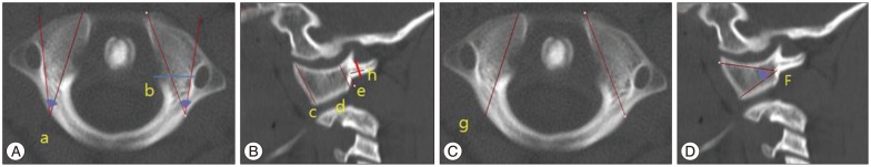
Fig. 1 Axial (A and C) and sagittal (B and D) computed tomographic images show the morphometric anatomy for measuring C1 cervical spine. a : medio-lateral angle, b : lateral mass width, c : lateral mass height (anterior), d : lateral mass height (posterior), e : entry point height, f : supero-inferior angle, g : maximal screw length, h : C1 posterior arch height.
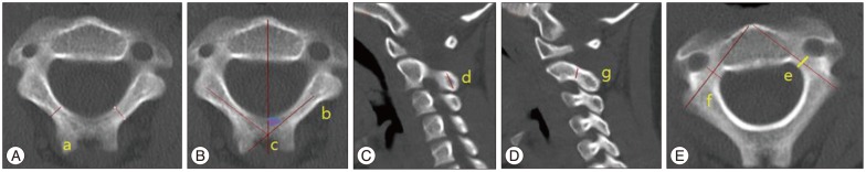
Fig. 2 Axial (A, B, and E) and sagittal (C and D) computed tomographic images show the morphometric anatomy for measuring C2 cervical spine. a : lamina width, b : lamina length, c : spino-lamina angle, d : lamina height, e : pedicle width, f : pedicle length, g : pedicle height.

Fig. 3 Axial (A, C, and E) and sagittal (B and D) computed tomographic images show the morphometric anatomy for measuring the subaxial spine. a : pedicle width, b : pedicle height, c : lamina width, d : lamina height, e : leteral mass length.

Fig. 4 Graph demonstrating the mean value of pedicle and lamina dimensions in subaxial spine. PW : pedicle width, PH : pedicle height, LW : lamina width, LH : lamina height.
Reference
-
1. Abumi K, Itoh H, Taneichi H, Kaneda K. Transpedicular screw fixation for traumatic lesions of the middle and lower cervical spine : description of the techniques and preliminary report. J Spinal Disord. 1994; 7:19–28. PMID: 8186585.
Article2. Anderson RC, Ragel BT, Mocco J, Bohman LE, Brockmeyer DL. Selection of a rigid internal fixation construct for stabilization at the craniovertebral junction in pediatric patients. J Neurosurg. 2007; 107(1 Suppl):36–42. PMID: 17644919.
Article3. Chamoun RB, Relyea KM, Johnson KK, Whitehead WE, Curry DJ, Luerssen TG, et al. Use of axial and subaxial translaminar screw fixation in the management of upper cervical spinal instability in a series of 7 children. Neurosurgery. 2009; 64:734–739. discussion 739. PMID: 19349831.
Article4. Chamoun RB, Whitehead WE, Curry DJ, Luerssen TG, Jea A. Computed tomography morphometric analysis for C-1 lateral mass screw placement in children. Clinical article. J Neurosurg Pediatr. 2009; 3:20–23. PMID: 19119899.
Article5. Chern JJ, Chamoun RB, Whitehead WE, Curry DJ, Luerssen TG, Jea A. Computed tomography morphometric analysis for axial and subaxial translaminar screw placement in the pediatric cervical spine. J Neurosurg Pediatr. 2009; 3:121–128. PMID: 19278311.
Article6. Cristante AF, Torelli AG, Kohlmann RB, Dias da, Biraghi OL, Iu-taka AS, et al. Feasibility of intralaminar, lateral mass, or pedicle axis vertebra screws in children under 10 years of age : a tomographic study. Neurosurgery. 2012; 70:835–838. discussion 838-839. PMID: 21937932.7. Ferri-de-Barros F, Little DG, Bridge C, Cummine J, Cree AK. Atlantoaxial and craniocervical arthrodesis in children : a tomographic study comparing suitability of C2 pedicles and C2 laminae for screw fixation. Spine (Phila Pa 1976). 2010; 35:291–293. PMID: 20075784.8. Grob D, Crisco JJ 3rd, Panjabi MM, Wang P, Dvorak J. Biomechanical evaluation of four different posterior atlantoaxial fixation techniques. Spine (Phila Pa 1976). 1992; 17:480–490. PMID: 1621145.
Article9. Hedequist D, Proctor M, Hresko T. Lateral mass screw fixation in children. J Child Orthop. 2010; 4:197–201. PMID: 21629379.
Article10. Hong JT, Sung JH, Son BC, Lee SW, Park CK. Significance of laminar screw fixation in the subaxial cervical spine. Spine (Phila Pa 1976). 2008; 33:1739–1743. PMID: 18628706.
Article11. Hong JT, Yi JS, Kim JT, Ji C, Ryu KS, Park CK. Clinical and radiologic outcome of laminar screw at C2 and C7 for posterior instrumentation--review of 25 cases and comparison of C2 and C7 intralaminar screw fixation. World Neurosurg. 2010; 73:112–118. discussion e15. PMID: 20860937.
Article12. Hwang SW, Gressot LV, Rangel-Castilla L, Whitehead WE, Curry DJ, Bollo RJ, et al. Outcomes of instrumented fusion in the pediatric cervical spine. J Neurosurg Spine. 2012; 17:397–409. PMID: 22998404.
Article13. Jea A, Johnson KK, Whitehead WE, Luerssen TG. Translaminar screw fixation in the subaxial pediatric cervical spine. J Neurosurg Pediatr. 2008; 2:386–390. PMID: 19035682.
Article14. Jea A, Taylor MD, Dirks PB, Kulkarni AV, Rutka JT, Drake JM. Incorporation of C-1 lateral mass screws in occipitocervical and atlantoaxial fusions for children 8 years of age or younger. Technical note. J Neurosurg. 2007; 107(2 Suppl):178–183. PMID: 18459894.
Article15. Jones EL, Heller JG, Silcox DH, Hutton WC. Cervical pedicle screws versus lateral mass screws. Anatomic feasibility and biomechanical comparison. Spine (Phila Pa 1976). 1997; 22:977–982. PMID: 9152447.16. Kanna PR, Shetty AP, Rajasekaran S. Anatomical feasibility of pediatric cervical pedicle screw insertion by computed tomographic morphometric evaluation of 376 pediatric cervical pedicles. Spine (Phila Pa 1976). 2011; 36:1297–1304. PMID: 21289586.
Article17. Leonard JR, Wright NM. Pediatric atlantoaxial fixation with bilateral, crossing C-2 translaminar screws. Technical note. J Neurosurg. 2006; 104(1 Suppl):59–63. PMID: 16509484.
Article18. Mandel IM, Kambach BJ, Petersilge CA, Johnstone B, Yoo JU. Morphologic considerations of C2 isthmus dimensions for the placement of transarticular screws. Spine (Phila Pa 1976). 2000; 25:1542–1547. PMID: 10851104.
Article19. Melcher RP, Puttlitz CM, Kleinstueck FS, Lotz JC, Harms J, Bradford DS. Biomechanical testing of posterior atlantoaxial fixation techniques. Spine (Phila Pa 1976). 2002; 27:2435–2440. PMID: 12435971.
Article20. Meng XZ, Xu JX. The options of C2 fixation for os odontoideum : a radiographic study for the C2 pedicle and lamina anatomy. Eur Spine J. 2011; 20:1921–1927. PMID: 21725866.
Article21. Oda I, Abumi K, Sell LC, Haggerty CJ, Cunningham BW, McAfee PC. Biomechanical evaluation of five different occipito-atlanto-axial fixation techniques. Spine (Phila Pa 1976). 1999; 24:2377–2382. PMID: 10586464.
Article22. Panjabi MM, Duranceau J, Goel V, Oxland T, Takata K. Cervical human vertebrae. Quantitative three-dimensional anatomy of the middle and lower regions. Spine (Phila Pa 1976). 1991; 16:861–869. PMID: 1948369.
Article23. Rajasekaran S, Kanna PR, Shetty AP. Safety of cervical pedicle screw insertion in children : a clinicoradiological evaluation of computer-assisted insertion of 51 cervical pedicle screws including 28 subaxial pedicle screws in 16 children. Spine (Phila Pa 1976). 2012; 37:E216–E223. PMID: 21912324.24. Sekhon LH. Posterior cervical lateral mass screw fixation : analysis of 1026 consecutive screws in 143 patients. J Spinal Disord Tech. 2005; 18:297–303. PMID: 16021008.
- Full Text Links
-
- Actions
-
Cited
- CITED
-
- Close
- Share
-
- Similar articles
-
- Cervical Pedicle Screw Fixation: Anatomic Feasibility of Pedicle Morphology and Radiologic Evaluation of the Anatomical Measurements
- Morphometric Study of C1 Pedicle and Feasibility Evaluation of C1 Pedicle Screw Placement with a Novel Clinically Relevant Radiological Classification in an Indian Population
- Patient-Specific Drill Guide Template for Pedicle Screw Insertion into the Atlantoaxial Cervical Spine Using Stereolithographic Modeling: An In Vitro Study
- C2 Anatomy for Translaminar Screw Placement Based on Computerized Tomographic Measurements
- Posterior Atlantoaxial Fixation with a Combination of Pedicle Screws and a Laminar Screw in the Axis for a Unilateral High-riding Vertebral Artery
