Chronic Encapsulated Intracerebral Hematoma Associated with Cavernous Malformation
- Affiliations
-
- 1Department of Neurosurgery, National Defense Medical College, Saitama, Japan. s.takeuchi@room.ocn.ne.jp
- KMID: 2191051
- DOI: http://doi.org/10.3340/jkns.2014.55.2.89
Abstract
- Chronic encapsulated intracerebral hematoma (CEIH) is a rare cerebrovascular disease that behaves as a slowly expanding lesion with a gradual onset. It is well established that CEIH is associated with arteriovenous malformations; however, CEIH associated with cavernous malformation (CM) is extremely rare. We herein report a case of CEIH associated with CM, and discuss its pathogenesis. A 12-year-old female was admitted to our hospital because of a one week history of progressive headache and nausea. Brain computed tomography scan and magnetic resonance imaging showed an intracerebral hematoma surrounded by edema in the right frontal lobe. One week later, her headache and nausea worsened, and a brain computed tomography scan revealed the enlargement of hematoma. A right frontal craniotomy was performed. The capsule, mass, and hematoma were totally removed. Histological examination confirmed the diagnosis of CEIH associated with CM. Immunohistochemical analysis revealed increased expression of vascular endothelial growth factor (VEGF) and the VEGF receptor-1 in the endothelium and fibroblasts. Our findings suggest that the activated VEGF pathway might have positively contributed to development of CEIH in the present patient.
Keyword
MeSH Terms
Figure
-
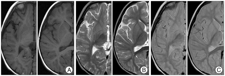
Fig. 1 Imaging findings at 21 months of age. Axial T1-weighted (A), T2-weighted (B), and proton density (C) magnetic resonance images show no evidence of any lesions.
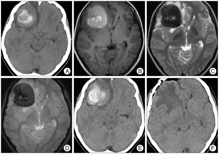
Fig. 2 Imaging findings during admission. A : Computed tomography (CT) scan on admission shows an intracerebral hematoma surrounded by edema in the right frontal lobe. B, C, and D : Axial T1-weighted (B), T2-weighted (C), and T2*-weighted (D) MRI reveal a mass lesion as mixed intensities. E : CT scan obtained one week after admission shows progression of hematoma. F : Postoperative CT scan shows total removal of the lesion.
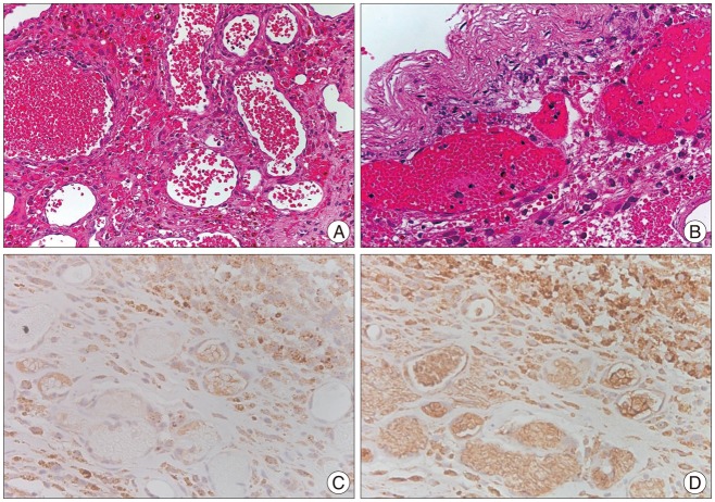
Fig. 3 Histopathological findings. A : Specimen (H&E) taken from the lobulated mass shows thin-walled dilated vascular channels with no intervening brain parenchyma, thus suggesting the presence of cavernous malformation. B : Specimen (H&E) of the capsule shows its structure consisting of an outer collagenous layer and an inner granulated layer with deposits of hemosiderin, which is compatible with chronic encapsulated intracerebral hematoma. C : Immunohistochemistry for vascular endothelial growth factor (VEGF) reveals positive immunostaining in the endothelium and fibroblasts. Original magnification : 400×. D : Immunohistochemistry for VEGF receptor-1 reveals positive immunostaining in the endothelium and fibroblasts. Original magnification : 400×.
Reference
-
1. Abe T, Morishige M, Ooba H, Kamida T, Fujiki M, Kobayashi H, et al. The association between high VEGF levels and multiple probable punctuate cavernous malformations. Acta Neurochir (Wien). 2009; 151:855–859. PMID: 19479188.
Article2. Cakirer S. De novo formation of a cavernous malformation of the brain in the presence of a developmental venous anomaly. Clin Radiol. 2003; 58:251–256. PMID: 12639533.
Article3. Campeau NG, Lane JI. De novo development of a lesion with the appearance of a cavernous malformation adjacent to an existing developmental venous anomaly. AJNR Am J Neuroradiol. 2005; 26:156–159. PMID: 15661718.4. Garner TB, Del Curling O Jr, Kelly DL Jr, Laster DW. The natural history of intracranial venous angiomas. J Neurosurg. 1991; 75:715–722. PMID: 1919693.
Article5. Jung KH, Chu K, Jeong SW, Park HK, Bae HJ, Yoon BW. Cerebral cavernous malformations with dynamic and progressive course : correlation study with vascular endothelial growth factor. Arch Neurol. 2003; 60:1613–1618. PMID: 14623736.
Article6. Kim YS, Lee JI, Choi CH, Ko JK. Massive intracerebral hemorrhage caused by a cavernous malformation. J Korean Neurosurg Soc. 2012; 51:37–39. PMID: 22396841.
Article7. Kondziolka D, Lunsford LD, Kestle JR. The natural history of cerebral cavernous malformations. J Neurosurg. 1995; 83:820–824. PMID: 7472549.
Article8. Massa-Micon B, Luparello V, Bergui M, Pagni CA. De novo cavernoma case report and review of literature. Surg Neurol. 2000; 53:484–487. PMID: 10874148.
Article9. Masuzawa T, Saito K, Shimabukuro H, Iwasa H, Sato F. Chronic encapsulated hematomas in the brain. Acta Neuropathol. 1985; 66:24–28. PMID: 3993333.
Article10. Miyahara K, Fujitsu K, Yagishita S, Ichikawa T, Takemoto Y, Okada T, et al. Chronic encapsulated intracerebral hematoma associated with cavernous angioma--case report. Neurol Med Chir (Tokyo). 2011; 51:52–55. PMID: 21273746.
Article11. Motegi H, Kuroda S, Ishii N, Aoyama H, Terae S, Shirato H, et al. De novo formation of cavernoma after radiosurgery for adult cerebral arteriovenous malformation--case report. Neurol Med Chir (Tokyo). 2008; 48:397–400. PMID: 18812682.
Article12. Murakami S, Sotsu M, Morooka S, Suzuki T. Chronic encapsulated intracerebral hematoma associated with cavernous angioma : a case report. Neurosurgery. 1990; 26:700–702. PMID: 2330096.
Article13. Nakamizo A, Suzuki SO, Saito N, Shono T, Matsumoto K, Onaka S, et al. Clinicopathological study on chronic encapsulated expanding hematoma associated with incompletely obliterated AVM after stereotactic radiosurgery. Acta Neurochir (Wien). 2011; 153:883–893. PMID: 20931239.
Article14. Roda JM, Carceller F, Pérez-Higueras A, Morales C. Encapsulated intracerebral hematomas : a defined entity. Case report. J Neurosurg. 1993; 78:829–833. PMID: 8468616.15. Takeuchi S, Nawashiro H, Wada K, Minamimura K. Chronic encapsulated intracerebral hematoma associated with cavernous angioma : expression of vascular endothelial growth factor and its receptor. Neurol India. 2011; 59:903–904. PMID: 22234209.
Article16. Takeuchi S, Takasato Y. Chronic encapsulated intracerebral hematoma : a rare complication after stereotactic radiosurgery for cerebral arteriovenous malformation. Acta Neurochir (Wien). 2011; 153:895. PMID: 21052741.
Article17. Takeuchi S, Takasato Y, Masaoka H. Chronic encapsulated intracerebral hematoma formation after radiosurgery for cerebral arteriovenous malformation. Neurol India. 2011; 59:624–626. PMID: 21891948.
Article18. Takeuchi S, Takasato Y, Masaoka H, Hayakawa T, Otani N, Yoshino Y, et al. Development of chronic encapsulated intracerebral hematoma after radiosurgery for a cerebral arteriovenous malformation. Acta Neurochir (Wien). 2009; 151:1513–1515. PMID: 19597762.
Article
- Full Text Links
-
- Actions
-
Cited
- CITED
-
- Close
- Share
-
- Similar articles
-
- Massive Intracerebral Hemorrhage Caused by a Cavernous Malformation
- Dural Arteriovenous Malformation on the Anterior Cranial Fossa
- Chronic Encapsulated Intracerebral Hematomas: So Called Chocolate Cysts
- A Neonatal Arteriovenous Malformation with Subdural and Intracerebral Hematoma
- Chronic Encapsulated Intracerebellar Hematoma Simulating Brain Tumor during Infancy
