Study on the position of the posterior superior alveolar artery in relation to the performance of the maxillary sinus bone graft procedure in a Korean population
- Affiliations
-
- 1Department of Oral and Maxillofacial Surgery, School of Dentistry, Kyungpook National University, Daegu, Korea. kimcs@knu.ac.kr
- KMID: 2189690
- DOI: http://doi.org/10.5125/jkaoms.2012.38.2.71
Abstract
OBJECTIVES
This study sought to investigate the positioning of the posterior superior alveolar artery in relation to the performance of the maxillary sinus bone graft procedure in a Korean population.
MATERIALS AND METHODS
We identified the position of the posterior superior alveolar artery relative to 93 maxillary sinuses in 58 patients and determined the distance from the inferior border of the artery in the premolar and molar areas to the alveolar ridge and sinus floor.
RESULTS
The mean distance from the alveolar ridge to the posterior superior alveolar artery in the dentate group (20.62+/-3.05 mm in the premolar region, 17.50+/-2.84 mm in the molar region) was greater than as compared to the edentulous group (18.83+/-2.79 mm in the premolar region, 15.50+/-1.64 mm in the molar region), and this difference was statistically significant (P<0.05). In contrast, there was no statistically significant difference (P>0.05) between the mean distance from the sinus floor to the posterior superior alveolar artery in the dentate group (8.21+/-2.79 mm in the premolar region, 7.52+/-2.07 mm in the molar region) or in the edentulous group (7.75+/-3.31 mm in the premolar region, 7.97+/-2.31 mm in the molar region).
CONCLUSION
Prior to surgery, it is important to evaluate the position of the posterior superior maxillary artery by using computed tomography scans. The premolar area is safer than the molar area for performing the maxillary sinus bone graft without bleeding.
MeSH Terms
Figure
-
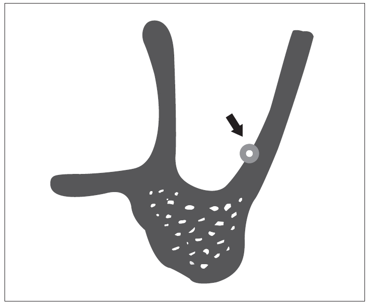
Fig. 1. Schematic representation of the cross-sectional view of the maxillary sinus. The bone canal is intraosseous anastomosis from the posterior superior alveolar artery (arrow indicates intraosseous anastomosis).
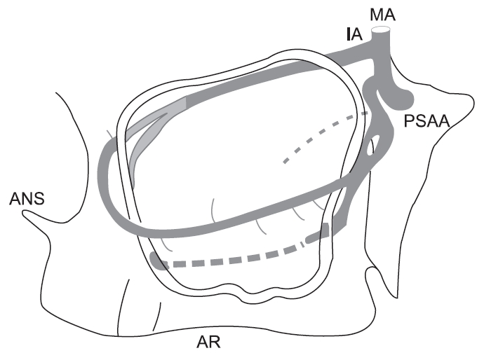
Fig. 2. Schematic representation of sagittal section of the maxillary sinus with blood vessels. (MA: maxillary artery, IA: infra-orbital artery, PSAA: posterior superior alveolar artery, ANS: anterior nasal spine, AR: alveolar ridge)
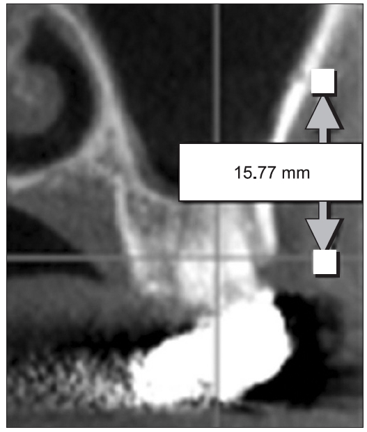
Fig. 3. Measurement of the distance from the alveolar ridge to the posterior superior alveolar artery on the computed tomography image.
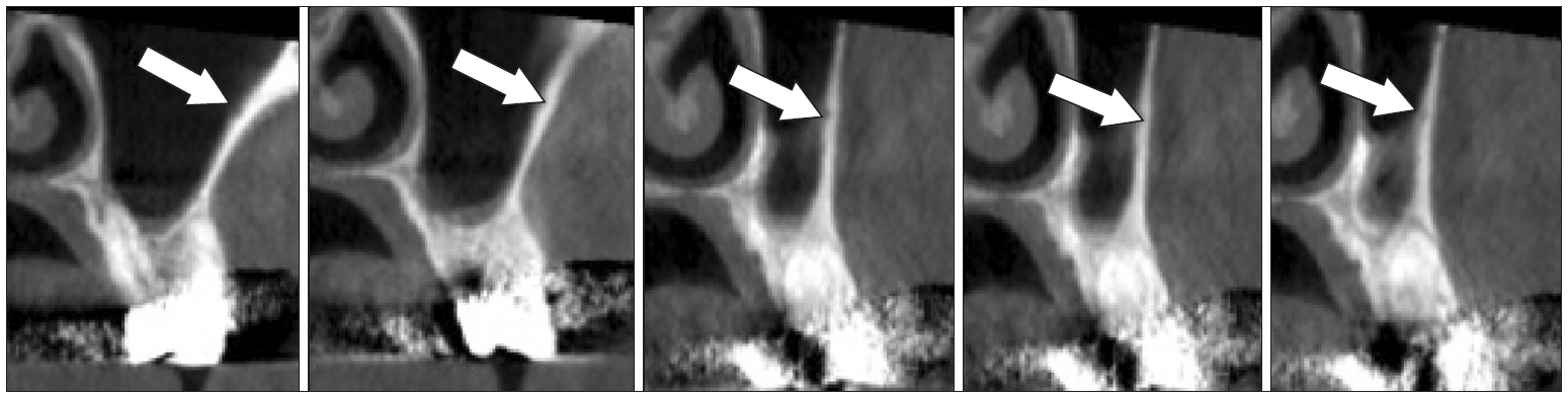
Fig. 4. Continuity of the posterior superior alveolar artery on the computed tomography T image.
Cited by 2 articles
-
Buccal infiltration injection without a 4% articaine palatal injection for maxillary impacted third molar surgery
Som Sochenda, Chakorn Vorakulpipat, Kumar K C, Chavengkiat Saengsirinavin, Manus Rojvanakarn, Natthamet Wongsirichat
J Korean Assoc Oral Maxillofac Surg. 2020;46(4):250-257. doi: 10.5125/jkaoms.2020.46.4.250.Cone-beam computed tomography characterization of the intraosseous vascular canal in the lateral wall of the maxillary antrum
Shishir Ram Shetty, Saad Wahby Al Bayatti, Hesham Marei, Raghavendra Shetty, Hossam Abdelatty Abdelmagyd, Alexander Maniangat Luke
J Korean Assoc Oral Maxillofac Surg. 2021;47(1):34-39. doi: 10.5125/jkaoms.2021.47.1.34.
Reference
-
1. Marcus SE, Drury TF, Brown LJ, Zion GR. Tooth retention and tooth loss in the permanent dentition of adults: United States, 1988-1991. J Dent Res. 1996; 75:684–695.
Article2. Pietrokovski J. The bony residual ridge in man. J Prosthet Dent. 1975; 34:456–462.
Article3. Jaffin RA, Berman CL. The excessive loss of Branemark fixtures in type IV bone: a 5-year analysis. J Periodontol. 1991; 62:2–4.
Article4. Shackleton JL, Carr L, Slabbert JC, Becker PJ. Survival of fixed implant-supported prostheses related to cantilever lengths. J Prosthet Dent. 1994; 71:23–26.
Article5. Choi J, Park HS. The clinical anatomy of the maxillary artery in the pterygopalatine fossa. J Oral Maxillofac Surg. 2003; 61:72–78.
Article6. Smiler DG, Johnson PW, Lozada JL, Misch C, Rosenlicht JL, Tatum OH Jr, et al. Sinus lift grafts and endosseous implants. Treatment of the atrophic posterior maxilla. Dent Clin North Am. 1992; 36:151–186.7. Jensen OT, Shulman LB, Block MS, Iacono VJ. Report of the Sinus Consensus Conference of 1996. Int J Oral Maxillofac Implants. 1998; 13 Suppl:11–45.8. Del Fabbro M, Testori T, Francetti L, Weinstein R. Systematic review of survival rates for implants placed in the grafted maxillary sinus. Int J Periodontics Restorative Dent. 2004; 24:565–577.
Article9. Olson JW, Dent CD, Morris HF, Ochi S. Long-term assessment (5 to 71 months) of endosseous dental implants placed in the augmented maxillary sinus. Ann Periodontol. 2000; 5:152–156.
Article10. Tong DC, Rioux K, Drangsholt M, Beirne OR. A review of survival rates for implants placed in grafted maxillary sinuses using meta-analysis. Int J Oral Maxillofac Implants. 1998; 13:175–182.11. The Korean Association of Oral and Maxillofacial Surgeons. Textbook of oral maxillofacial surgery. 2nd ed. Seoul: Uichihaksa; 2005. pp. 195.12. Ikeda A, Ikeda M, Komatsuzaki A. A CT study of the course of growth of the maxillary sinus: normal subjects and subjects with chronic sinusitis. ORL J Otorhinolaryngol Relat Spec. 1998; 60:147–152.
Article13. Uchida Y, Goto M, Katsuki T, Soejima Y. Measurement of maxillary sinus volume using computerized tomographic images. Int J Oral Maxillofac Implants. 1998; 13:811–818.14. Barone A, Santini S, Sbordone L, Crespi R, Covani U. A clinical study of the outcomes and complications associated with maxillary sinus augmentation. Int J Oral Maxillofac Implants. 2006; 21:81–85.15. Tatum H Jr. Maxillary and sinus implant reconstructions. Dent Clin North Am. 1986; 30:207–229.16. Summers RB. A new concept in maxillary implant surgery: the osteotome technique. Compendium. 1994; 15:152, 154–6, 158 passim. quiz 162.17. Rosen PS, Summers R, Mellado JR, Salkin LM, Shanaman RH, Marks MH, et al. The bone-added osteotome sinus floor elevation technique: multicenter retrospective report of consecutively treated patients. Int J Oral Maxillofac Implants. 1999; 14:853–858.18. Toffler M. Osteotome-mediated sinus floor elevation: a clinical report. Int J Oral Maxillofac Implants. 2004; 19:266–273.19. van den Bergh JP, ten Bruggenkate CM, Disch FJ, Tuinzing DB. Anatomical aspects of sinus floor elevations. Clin Oral Implants Res. 2000; 11:256–265.
Article20. Traxler H, Windisch A, Geyerhofer U, Surd R, Solar P, Firbas W. Arterial blood supply of the maxillary sinus. Clin Anat. 1999; 12:417–421.
Article21. Solar P, Geyerhofer U, Traxler H, Windisch A, Ulm C, Watzek G. Blood supply to the maxillary sinus relevant to sinus floor elevation procedures. Clin Oral Implants Res. 1999; 10:34–44.
Article22. Elian N, Wallace S, Cho SC, Jalbout ZN, Froum S. Distribution of the maxillary artery as it relates to sinus floor augmentation. Int J Oral Maxillofac Implants. 2005; 20:784–787.23. Mardinger O, Abba M, Hirshberg A, Schwartz-Arad D. Prevalence, diameter and course of the maxillary intraosseous vascular canal with relation to sinus augmentation procedure: a radiographic study. Int J Oral Maxillofac Surg. 2007; 36:735–738.
Article24. Kim KY, Kim SG, Seo HS, Song YJ, Kim MJ, Hong SM, et al. Arterial arcade of the maxillary sinus related to sinus bone graft in korean population ; A preliminary study using computed topographies. J Korean Assoc Oral Maxillofac Surg. 2008; 34:475–479.25. Jung YH, Nah KS, Cho BH. Maxillary sinus pneumatization after maxillary molar extraction assessed with cone beam computed tomography. Korean J Oral Maxillofac Radiol. 2009; 39:109–113.26. Sharan A, Madjar D. Maxillary sinus pneumatization following extractions: a radiographic study. Int J Oral Maxillofac Implants. 2008; 23:48–56.
- Full Text Links
-
- Actions
-
Cited
- CITED
-
- Close
- Share
-
- Similar articles
-
- The literature review on the sinus bone graft using deproteinized bovine bone mineral with lateral approach
- SINUS GRAFT AND VERTICAL AUGMENTATION OF MAXILLARY POSTERIOR ALVEOLAR RIDGE USING MANDIBULAR RAMAL BLOCK BONE GRAFT
- Vertical Augmentation of Maxillary Posterior Alveolar Ridge Using Allogenic Block Bone Graft and Simultaneous Maxillary Sinus Graft
- Evaluation of the posterior superior alveolar artery canal by cone-beam computed tomography in a sample of the Egyptian population
- Arterial arcade of the maxillary sinus related to sinus bone graft in korean population ; A preliminary study using computed topographies
