J Korean Assoc Oral Maxillofac Surg.
2015 Apr;41(2):78-83. 10.5125/jkaoms.2015.41.2.78.
Mandibular intraosseous squamous cell carcinoma lesion associated with odontogenic keratocyst: a case report
- Affiliations
-
- 1Department of Oral and Maxillofacial Surgery, School of Dentistry, Pusan National University, Yangsan, Korea. kuksjs@pusan.ac.kr
- 2Department of Oral and Maxillofacial Surgery, Ulsan University Hospital, Ulsan, Korea.
- KMID: 2189467
- DOI: http://doi.org/10.5125/jkaoms.2015.41.2.78
Abstract
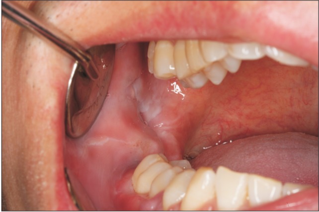
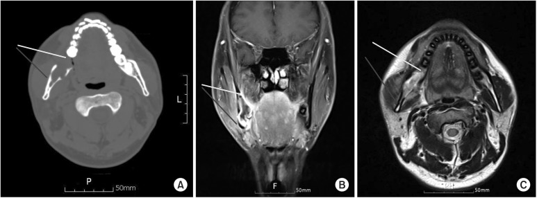
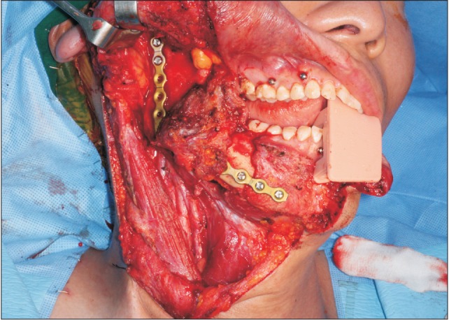
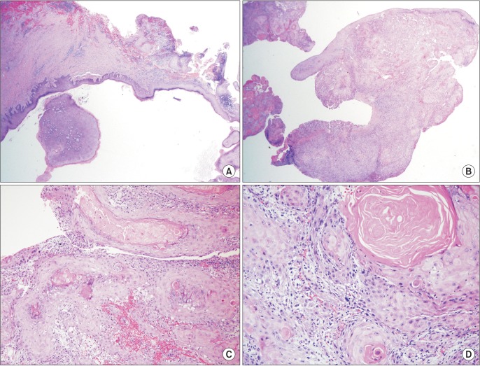
- Squamous cell carcinoma (SCC) is the most common malignant tumor in the oral cavity, and it accounts for about 90% of all oral cancers. Several risk factors for oral SCC have been identified; however, SCC associated with odontogenic keratocysts have rarely been reported. The present study describes the case of a 36-year-old man with SCC of the right ramus of the mandible, which was initially diagnosed as a benign odontogenic cyst. He underwent enucleation at another hospital followed by segmental mandibulectomy and fibular free flap reconstruction at our institution. In this case, we introduce a patient with oral cancer associated with odontogenic cyst on the mandible and report a satisfactory outcome with wide resection and immediate free flap reconstruction.
MeSH Terms
Figure
Cited by 1 articles
-
Sequential treatment from mandibulectomy to reconstruction on mandibular oral cancer – Case review II: mandibular anterior and the floor of the mouth lesion of basaloid squamous cell carcinoma and clear cell odontogenic carcinoma
Jae-Young Yang, Dae-Seok Hwang, Uk-Kyu Kim
J Korean Assoc Oral Maxillofac Surg. 2021;47(3):216-223. doi: 10.5125/jkaoms.2021.47.3.216.
Reference
-
1. Mohtasham N, Babazadeh F, Jafarzadeh H. Intraosseous verrucous carcinoma originating from an odontogenic cyst: a case report. J Oral Sci. 2008; 50:91–94. PMID: 18403890.
Article2. Marx RE, Stern D. Oral and maxillofacial pathology: a rationale for diagnosis and treatment. Hanover Park: Quintessence Publishing;2012.3. Shafer WG, Hine MK, Levy BN. A textbook of oral pathology. 4th ed. Philadelphia: W.B. Saunders;1983. p. 271–273.4. Loos D. Central epidermoid carcinoma of the jaws. Dtsch Monatsschr Zahnheilk. 1913; 31:308.5. Schwimmer AM, Aydin F, Morrison SN. Squamous cell carcinoma arising in residual odontogenic cyst. Report of a case and review of literature. Oral Surg Oral Med Oral Pathol. 1991; 72:218–221. PMID: 1923401.6. Keszler A, Piloni MJ. Malignant transformation in odontogenic keratocysts. Case report. Med Oral. 2002; 7:331–335. PMID: 12415216.7. Tan B, Yan TS, Shermin L, Teck KC, Yoke PC, Goh C, et al. Malignant transformation of keratocystic odontogenic tumor: two case reports. Am J Otolaryngol. 2013; 34:357–361. PMID: 23374486.
Article8. Gardner AF. The odontogenic cyst as a potential carcinoma: A clinicopathologic appraisal. J Am Dent Assoc. 1969; 78:746–755. PMID: 4887224.
Article9. Bodner L, Manor E, Shear M, van der Waal I. Primary intraosseous squamous cell carcinoma arising in an odontogenic cyst: a clinicopathologic analysis of 116 reported cases. J Oral Pathol Med. 2011; 40:733–738. PMID: 21689161.10. Pindborg JJ, Kramer IRH, Torioni H. Histologic typing of odontogenic tumours, cysts, and allied lesions. Geneva: World Health Organization;1971. p. 35.11. Elzay RP. Primary intraosseous carcinoma of the jaws. Review and update of odontogenic carcinomas. Oral Surg Oral Med Oral Pathol. 1982; 54:299–303. PMID: 6957827.12. Waldron CA. Oral epithelial tumors. In : Thoma KH, Gorlin RJ, Goldman HM, editors. Thoma's oral pathology. 4th ed. St. Louis: Mosby;1970. p. 846–847.13. Shear M. Primary intra-alveolar epidermoid carcinoma of the jaw. J Path. 1969; 97:645–651. PMID: 5354042.
Article14. Bereket C, Bekçioğlu B, Koyuncu M, Sener I, Kandemir B, Türer A. Intraosseous carcinoma arising from an odontogenic cyst: a case report. Oral Surg Oral Med Oral Pathol Oral Radiol. 2013; 116:e445–e449. PMID: 22921445.
Article15. Areen RG, McClatchey KD, Baker HL. Squamous cell carcinoma developing in an odontogenic keratocyst. Arch Otolaryngol. 1981; 107:568–569. PMID: 7271558.
- Full Text Links
- Actions
-
Cited
- CITED
-
- Close
- Share
- Similar articles
-
- Squamous Cell Carcinoma of the Maxilla Originated in Odontogenic Cyst: A Case Report
- A Case of Squamous Cell Carcinoma arising from an Odontogenic Keratocyst
- Primary intraosseous carcinoma(PIOC) on mandible: Case Report
- Squamous cell carcinoma arising within a maxillary odontogenic keratocyst: A rare occurrence
- Sequential treatment from mandibulectomy to reconstruction on mandibular oral cancer – Case review I: mandibular ramus and angle lesion of primary intraosseous squamous cell carcinoma

