Three Dimensional Analysis for the Intramedullary Canal Axis of the Proximal Tibia: Clinical Relevance to Total Knee Arthroplasty
- Affiliations
-
- 1Department of Orthopedic Surgery, College of Medicine, Kyung Hee University, Seoul, Korea.
- 2Department of Orthopedic Surgery, College of Medicine, Hanyang University, Seoul, Korea. chhchoi@hanyang.ac.kr
- KMID: 2186559
- DOI: http://doi.org/10.4055/jkoa.2007.42.3.345
Abstract
-
PURPOSE: To evaluate the appropriate entry point of an intramedullary tibial cutting guide in total knee arthroplasty in Koreans by measuring the "intramedullary canal axis" of the proximal tibia in three dimensions.
MATERIALS AND METHODS
Computed tomography was performed on 116 lower extremities from the hip to the ankle on 58 Korean cadavers. A three dimensional image of the tibia was reconstructed using the program, Bionix version 3.3. The location of intramedullary canal axis of proximal tibia passing through tibial plateau, canal axis center 1 (CAC 1), was measured. The beta' angle was defined as the angle between the tibial anatomical axis and a line perpendicular to the knee joint line. The correlations between the beta' angle and the CAC 1 mediolateral coordinates were analyzed.
RESULTS
The CAC 1 is located on 56.3% from the medial cortex and 27.8% from the anterior cortex on the average. On average, the CAC 1 was located 1.2 mm medially and 15.9 mm anteriorly from the lateral tibial spine 1. The beta' angles and medial-lateral coordinates of the CAC 1 showed a significant positive correlation (r=0.418, p=0.000).
CONCLUSION
When using an intramedullary guide for tibial cutting in total knee arthroplasty in Koreans, the entry point at the lateral and anterior positions from the surface center of the tibial plateau is appropriate. The lateralization of the entry point of intramedullary tibial cutting guide becomes necessary as the varus of the tibia becomes more severe. Because of the marked variability in the CAC 1, a preoperative evaluation of the CAC 1 needs to be carried out in order to properly locate the appropriate entry point of the intramedullary tibial cutting guide in total knee arthroplasty.
MeSH Terms
Figure
-

Fig. 1 The surface involving the transepicondylar axis is projected in the superior perspective of the tibial plateau and in the imaginary cut surface 10 mm below the lateral tibial plateau.
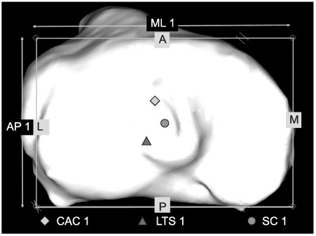
Fig. 2 Measured morphological parameters on the tibial plateau in the superior perspective. CAC 1, Canal axis center 1, is the location of intramedullary canal axis of the proximal tibia passing through the tibial plateau. LTS 1 is the location of the lateral tibial spine. SC 1, Surface center 1, is the anteroposterior and mediolateral center on the tibial plateau.
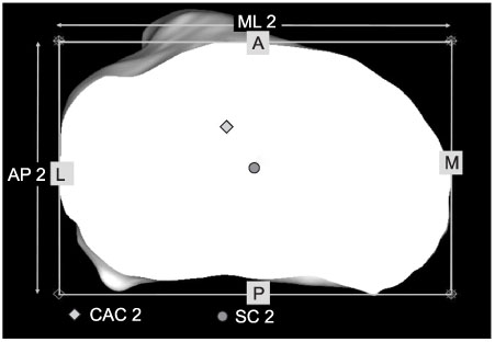
Fig. 3 Measured morphological parameters on the imaginary cut surface, 10 mm below the lateral tibial plateau in the superior perspective. CAC 2, Canal axis center 2, is the location of the intramedullary canal axis of the proximal tibia passing through an imaginary cut surface. SC 2, Surface center 2, is the anteroposterior and mediolateral center.
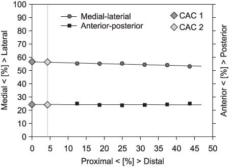
Fig. 4 Mathematically calculated canal axis center. The canal axis center was calculated using a least-squares fit to describe a line through the centroids.
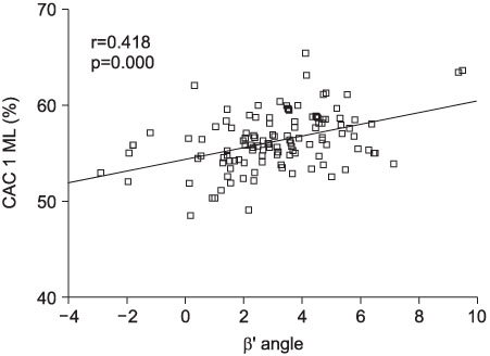
Fig. 5 Correlation between the β' angles and the CAC 1 mediolateral coordinates.
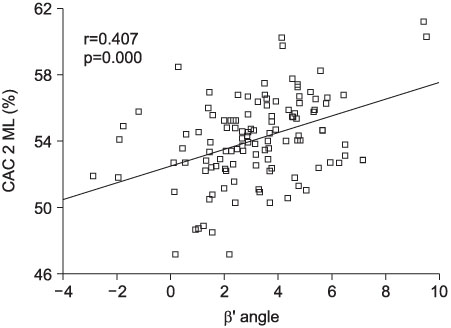
Fig. 6 Correlation between the β' angles and the CAC 2 mediolateral coordinates.
Reference
-
1. Bae DK, Park JY. The study of anatomical measurement of proximal tibia and fitness of tibial prosthesis in total knee arthroplasty. J Korean Orthop Assoc. 2000. 35:57–64.
Article2. Bargren JH, Blaha JD, Freeman MA. Alignment in total knee arthroplasty. Correlated biomechanical and clinical observations. Clin Orthop Relat Res. 1983. 173:178–183.3. Bartel DL, Burstein AH, Santavicca EA, Insall JN. Performance of the tibial component in total knee replacement. J Bone Joint Surg Am. 1982. 64:1026–1033.
Article4. Benjamin J. Component alignment in total knee arthroplasty. Instr Course Lect. 2006. 55:405–411.5. Bin SI, Choi JW, Nam TS. Total knee arthroplasty using intramedullary tibial cutting guide. J Korean Knee Soc. 2003. 15:17–21.6. Bindeiglass DF, Cohen JL, Dorr LD. Current principles of design for cemented and cementless knee. Tech Orthop. 1994. 6:80–85.7. Bloebaum RD, Bachus KN, Mitchell W, Hofmann G, Hofmann AA. Analysis of the bone surface area in resected tibia. Implications in tibial component subsidence and fixation. Clin Orthop Relat Res. 1994. 309:2–10.8. Bourne RB, Finlay JB, Papadopoulos P, Rorabeck CH, Andreae P. In vitro strain distribution in the proximal tibia. Effect of varus-valgus loading in the normal and osteoarthritic knee. Clin Orthop Relat Res. 1984. 188:285–291.9. Bourne RB, Finlay JB. The influence of tibial component intramedullary stems and implant-cortex contact on the strain distribution of the proximal tibia following total knee arthroplasty. An in vitro study. Clin Orthop Relat Res. 1986. 208:95–99.10. Brys DA, Lombardi AV Jr, Mallory TH, Vaughn BK. A comparison of intramedullary and extramedullary alignment systems for tibial component placement in total knee arthroplasty. Clin Orthop Relat Res. 1991. 263:175–179.
Article11. Cates HE, Ritter MA, Keating EM, Faris PM. Intramedullary versus extramedullary femoral alignment systems in total knee replacement. Clin Orthop Relat Res. 1993. 286:32–39.
Article12. Chung HK, Choi CH, Kim JH, Kim KT, Kim SI, Chang DP. Tibial plateau coverage in total knee replacement arthroplasty: coverage on 12 quadrants. J Korean Orthop Assoc. 1999. 34:1081–1086.
Article13. Dennis DA, Channer M, Susmann MH, Stringer EA. Intramedullary versus extramedullary tibial alignment systems in total knee arthroplasty. J Arthroplasty. 1993. 8:43–47.
Article14. Figgie HE 3rd, Davy DT, Heiple KG, Hart RT. Load-bearing capacity of the tibial component of the total condylar knee prosthesis An in vitro study. Clin Orthop Relat Res. 1984. 183:288–297.15. Hicks CA, Noble P, Tullos H. The anatomy of the tibial intramedullary canal. Clin Orthop Relat Res. 1995. 321:111–116.
Article16. Hofmann AA, Bachus KN, Wyatt RW. Effect of the tibial cut on subsidence following total knee arthroplasty. Clin Orthop Relat Res. 1991. 269:63–69.
Article17. Insall JN, Windsor RE, Scott WN. Surgical techniques and instrumentation in total knee arthroplasty. Surgery of the knee. 2001. 3rd ed. New York: Churchill Livingstone;1584–1587.18. Jeffcote B, Shakespeare D. Varus/valgus alignment of the tibial component in total knee arthroplasty. Knee. 2003. 10:243–247.
Article19. Kang SY, Lee EW, Kang KS, Lee HJ, Jung HJ, Jeong PH. Anatomical assessment of the proper insertion site for tibial nailing. J Korean Fracture Soc. 2004. 17:142–147.20. Lee RW, Volz RG, Sheridan DC. The role of fixation and bone quality on the mechanical stability of tibial knee components. Clin Orthop Relat Res. 1991. 273:177–183.
Article21. Maestro A, Harwin SF, Sandoval MG, Vaquero DH, Murcia A. Influence of intramedullary versus extramedullary alignment guides on final total knee arthroplasty component position: a radiographic analysis. J Arthroplasty. 1998. 5:552–558.22. Matsui Y, Kadoya Y, Uehara K, Kobayashi A, Takaoka K. Rotational deformity in varus osteoarthritis of the knee: analysis with computed tomography. Clin Orthop Relat Res. 2005. 433:147–151.23. Moreland JR. Mechanisms of failure in total knee arthroplasy. Clin Orthop Relat Res. 1988. 226:49–64.24. Nam WD, Rhyu KH, Han KY. The change of proximal reference of tibial plateau in total knee replacement: the cadaveric study. J Korean Knee Soc. 2005. 17:69–72.25. Ritter MA, Faris PM, Keating EM, Meding JB. Postoperative alignment of total knee replacement. Its effect on survival. Clin Orthop Relat Res. 1994. 299:153–156.
Article26. Robertson DD, Yuan J, Bigliani LU, Flatow EL, Yamaguchi K. Three-dimensional analysis of the proximal part of the humerus: relevance to arthroplasty. J Bone Joint Surg Am. 2000. 82:1594–1602.
Article27. Simmons ED Jr, Sullivan JA, Rackemann S, Scott RD. The accuracy of tibial intramedullary alignment devices in total knee arthroplasty. J Arthroplasty. 1991. 6:45–50.
Article28. Sokal RR, Rohlf FJ. Biometry. 1995. 3rd ed. New York: William H. Freeman;460–461. .29. Stern SH, Sharrock N, Kahn R, Insall JN. Haematologic and circulatory changes associated with total knee arthroplasty surgical instrumentation. Clin Orthop Realt Res. 1994. 299:179–189.30. Stulberg BN, Dombrowski RM, Froimson M, Easley K. Computed tomography analysis of proximal tibia coverage. Clin Orthop Relat Res. 1995. 311:148–156.31. Westrich GH, Haas SB, Insall JN, Frachie A. Resection specimen analysis of proximal tibial anatomy based on 100 total knee arthroplasty specimens. J Arthroplasty. 1995. 10:47–51.
Article32. Whiteside LA, Summers RG. Anatomical landmarks for an intramedullary alignment system for total knee replacement. Orthop Trans. 1983. 7:546–551.
- Full Text Links
-
- Actions
-
Cited
- CITED
-
- Close
- Share
-
- Similar articles
-
- Radiographic Analysis of the Tibial Axis on the Antero-posterior and Lateral view of Knee
- Tibial intramedullary canal axis and its influence on the intramedullary alignment system entry point in Koreans
- Total Knee Arthroplasty using Intramedullary Tibial Cutting Guide
- Accuracy of Intramedullary versus Extramedullary Tibial Alignment Guides: A Randomized, Prospective Study in Bilateral Total Knee Arthroplasty
- The Position of Proximal Reference Point of Tibial Plateau to Prevent Varus Tibial Cutting in Total Knee Replacement : Prospective Randomized Study
