J Gastric Cancer.
2014 Dec;14(4):252-258. 10.5230/jgc.2014.14.4.252.
Impact of Intraoperative Macroscopic Diagnosis of Serosal Invasion in Pathological Subserosal (pT3) Gastric Cancer
- Affiliations
-
- 1Department of Surgery, Yeouido St. Mary's Hospital, The Catholic University of Korea College of Medicine, Seoul, Korea. kimwook@catholic.ac.kr
- 2Department of Surgery, Bucheon St. Mary's Hospital, The Catholic University of Korea College of Medicine, Bucheon, Korea.
- KMID: 2183649
- DOI: http://doi.org/10.5230/jgc.2014.14.4.252
Abstract
- PURPOSE
The macroscopic diagnosis of tumor invasion through the serosa during surgery is not always distinct in patients with gastric cancer. The prognostic impact of the difference between macroscopic findings and pathological diagnosis of serosal invasion is not fully elucidated and needs to be re-evaluated.
MATERIALS AND METHODS
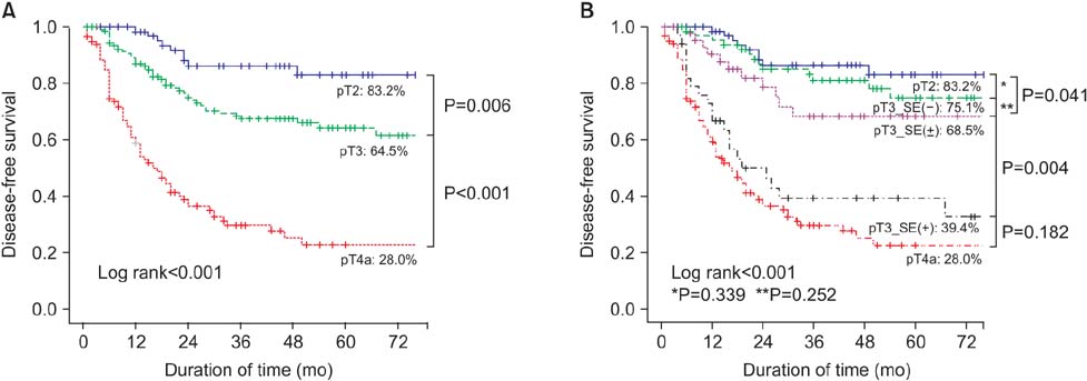
A total of 370 patients with locally advanced pT2 to pT4a gastric cancer who underwent curative surgery were enrolled in this study. Among them, 155 patients with pT3 were divided into three groups according to the intraoperative macroscopic diagnosis of serosal invasion, as follows: serosa exposure (SE)(-) (no invasion, 72 patients), SE(+/-) (ambiguous, 47 patients), and SE(+) (definite invasion, 36 patients), and the clinicopathological features, surgical outcomes, and disease-free survival (DFS) were analyzed.
RESULTS
A comparison of the 5-year DFS between pT3_SE(-) and pT2 groups and between pT3_SE(+) and pT4a groups revealed that the differences were not statistically significant. In addition, in a subgroup analysis of pT3 patients, the 5-year DFS was 75.1% in SE(-), 68.5% in SE(+/-), and 39.4% in SE(+) patients (P<0.05). In a multivariate analysis to evaluate risk factors for tumor recurrence, macroscopic diagnosis (hazard ratio [HR], SE(-) : SE(+/-) : SE(+)=1 : 1.01 : 2.45, P=0.019) and lymph node metastasis (HR, N0 : N1 : N2 : N3=1 : 1.45 : 2.20 : 9.82, P<0.001) were independent risk factors for recurrence.
CONCLUSIONS
Gross inspection of serosal invasion by the surgeon had a strong impact on tumor recurrence in gastric cancer patients. Consequently, the gross appearance of serosal invasion should be considered as a factor for predicting patients' prognosis.
Keyword
MeSH Terms
Figure
Reference
-
1. Jeong O, Ryu SY, Jeong MR, Sun JW, Park YK. Accuracy of macroscopic intraoperative diagnosis of serosal invasion and risk of over- and underestimation in gastric carcinoma. World J Surg. 2011; 35:2252–2258.
Article2. Korenaga D, Okuyama T, Orita H, Anai H, Baba H, Maehara Y, et al. Role of intraoperative assessment of lymph node metastasis and serosal invasion in patients with gastric cancer. J Surg Oncol. 1994; 55:250–254.
Article3. Ichiyoshi Y, Maehara Y, Tomisaki S, Oiwa H, Sakaguchi Y, Ohno S, et al. Macroscopic intraoperative diagnosis of serosal invasion and clinical outcome of gastric cancer: risk of underestimation. J Surg Oncol. 1995; 59:255–260.
Article4. Sobin LH, Gospodarowicz MK, Wittekind C, editors. International Union against Cancer. TNM Classification of Malignant Tumours. 7th ed. Chichester, West Sussex (UK), Hoboken (NJ): Wiley-Blackwell;2010.5. Japanese Gastric. Japanese classification of gastric carcinoma: 3rd English edition. Gastric Cancer. 2011; 14:101–112.
Article6. Kodera Y, Yamamura Y, Shimizu Y, Torii A, Hirai T, Yasui K, et al. Peritoneal washing cytology: prognostic value of positive findings in patients with gastric carcinoma undergoing a potentially curative resection. J Surg Oncol. 1999; 72:60–64. discussion 64-65.
Article7. La Torre M, Ferri M, Giovagnoli MR, Sforza N, Cosenza G, Giarnieri E, et al. Peritoneal wash cytology in gastric carcinoma. Prognostic significance and therapeutic consequences. Eur J Surg Oncol. 2010; 36:982–986.
Article8. Haraguchi M, Watanabe A, Kakeji Y, Tsujitani S, Baba H, Maehara Y, et al. Prognostic significance of serosal invasion in carcinoma of the stomach. Surg Gynecol Obstet. 1991; 172:29–32.9. Soga K, Ichikawa D, Yasukawa S, Kubota T, Kikuchi S, Fujiwara H, et al. Prognostic impact of the width of subserosal invasion in gastric cancer invading the subserosal layer. Surgery. 2010; 147:197–203.
Article10. Yasuda K, Shiraishi N, Inomata M, Shiroshita H, Izumi K, Kitano S. Prognostic significance of macroscopic serosal invasion in advanced gastric cancer. Hepatogastroenterology. 2007; 54:2028–2031.11. Giuliani A, Caporale A, Di Bari M, Demoro M, Gozzo P, Corona M, et al. Maximum gastric cancer diameter as a prognostic indicator: univariate and multivariate analysis. J Exp Clin Cancer Res. 2003; 22:531–538.12. Xu CY, Shen JG, Shen JY, Chen WJ, Wang LB. Ulcer size as a novel indicator marker is correlated with prognosis of ulcerative gastric cancer. Dig Surg. 2009; 26:312–316.
Article13. Song KY, Hur H, Jung CK, Jung ES, Kim SN, Jeon HM, et al. Impact of tumor infiltration pattern into the surrounding tissue on prognosis of the subserosal gastric cancer (pT2b). Eur J Surg Oncol. 2010; 36:563–567.
Article
- Full Text Links
- Actions
-
Cited
- CITED
-
- Close
- Share
- Similar articles
-
- Macroscopic Serosal Invasion in Advanced Gastric Cancer
- Prognostic significance of intraoperative macroscopic serosal invasion finding when it shows a discrepancy in pathologic result gastric cancer
- Usefulness of Spiral CT for T Staging of Gastric Carcinoma
- Significance of Carcinoembryonic Antigen Levels in Peritoneal Washings for Gastric Cancer Patients
- A study on double contrast study in early gastric cancer


