J Gynecol Oncol.
2012 Jan;23(1):69-71. 10.3802/jgo.2012.23.1.69.
Late presentation of metastatic smooth muscle neoplasm of the uterus with low malignant potential
- Affiliations
-
- 1University of East Anglia Medical School, Norwich, UK.
- 2Department of Obstetrics and Gynaecology, Norfolk and Norwich University Hospital NHS Foundation Trust, Norwich, UK. nikolaos.burbos@nnuh.nhs.uk
- 3Department of Histopathology, Norfolk and Norwich University Hospital NHS Foundation Trust, Norwich, UK.
- KMID: 2177517
- DOI: http://doi.org/10.3802/jgo.2012.23.1.69
Abstract
- A 48-year-old woman underwent total abdominal hysterectomy with conservation of the ovaries and tubes. Histology showed a well-circumscribed smooth muscle tumor with foci of degeneration (including infarct-type necrosis) but no coagulative tumor cell necrosis and only mild focal cytological atypia. She presented, 24 years later with shortness of breath and abdominal distension and underwent bilateral salpingo-oophorectomy, appendectomy, omental biopsy and para-aortic lymph node sampling. Histology showed bilateral ovarian smooth muscle tumors with no coagulative tumor cell necrosis or significant cellular atypia. The cells were mitotically active. The tumors in both ovaries were most likely secondary to the previous uterine smooth muscle neoplasm. To our knowledge, this case is the first in the literature to describe a benign cellular leiomyoma that subsequently behaved as a smooth muscle tumor of uncertain malignant potential, which recurred 24 years after the initial diagnosis.
MeSH Terms
Figure
-

Fig. 1 Cellular smooth muscle tumor with no significant cytological atypia (H&E, ×200).
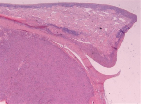
Fig. 2 Ovary showing well-circumscribed nodule of smooth muscle tumor (H&E, ×20).
Reference
-
1. Atkins KA, Arronte N, Darus CJ, Rice LW. The Use of p16 in enhancing the histologic classification of uterine smooth muscle tumors. Am J Surg Pathol. 2008. 32:98–102.2. Bell SW, Kempson RL, Hendrickson MR. Problematic uterine smooth muscle neoplasms: a clinicopathologic study of 213 cases. Am J Surg Pathol. 1994. 18:535–558.3. Berretta R, Rolla M, Merisio C, Giordano G, Nardelli GB. Uterine smooth muscle tumor of uncertain malignant potential: a three-case report. Int J Gynecol Cancer. 2008. 18:1121–1126.4. Guntupalli SR, Ramirez PT, Anderson ML, Milam MR, Bodurka DC, Malpica A. Uterine smooth muscle tumor of uncertain malignant potential: a retrospective analysis. Gynecol Oncol. 2009. 113:324–326.5. D'Angelo E, Prat J. Uterine sarcomas: a review. Gynecol Oncol. 2010. 116:131–139.6. Shapiro A, Ferenczy A, Turcotte R, Bruchim I, Gotlieb WH. Uterine smooth-muscle tumor of uncertain malignant potential metastasizing to the humerus as a high-grade leiomyosarcoma. Gynecol Oncol. 2004. 94:818–820.7. Amant F, Moerman P, Vergote I. Report of an unusual problematic uterine smooth muscle neoplasm, emphasizing the prognostic importance of coagulative tumor cell necrosis. Int J Gynecol Cancer. 2005. 15:1210–1212.8. Ip PP, Tse KY, Tam KF. Uterine smooth muscle tumors other than the ordinary leiomyomas and leiomyosarcomas: a review of selected variants with emphasis on recent advances and unusual morphology that may cause concern for malignancy. Adv Anat Pathol. 2010. 17:91–112.9. Ng JS, Han A, Chew SH, Low J. A clinicopathologic study of uterine smooth muscle tumors of uncertain malignant potential (STUMP). Ann Acad Med Singapore. 2010. 39:625–628.10. Ip PP, Cheung AN, Clement PB. Uterine smooth muscle tumors of uncertain malignant potential (STUMP): a clinicopathologic analysis of 16 cases. Am J Surg Pathol. 2009. 33:992–1005.
- Full Text Links
-
- Actions
-
Cited
- CITED
-
- Close
- Share
-
- Similar articles
-
- A Case of Cutaneous Smooth Muscle Tumor of Uncertain Malignant Potential
- A Case of Leiomyosarcoma of the Tongue
- A case of retroperitoneal huge smooth muscle tumor misleading to ovarian cancer
- Transvaginal Color Doppler Sonographv of Uterine Smooth Muscle Tumors: Prediction of the Extent of Degenerative Change and Differentiation of Leiomyosarcoma from Myoma
- Metastatic malignant melanoma with peritoneal seeding in a young woman: A case report
