J Cardiovasc Ultrasound.
2014 Mar;22(1):40-42. 10.4250/jcu.2014.22.1.40.
Multiple Papillary Fibroelastomas and Thrombus in the Left Heart
- Affiliations
-
- 1Division of Cardiology, Department of Internal Medicine, Haeundae Paik Hospital, Inje University College of Medicine, Busan, Korea. hacemed@hanmail.net
- 2Department of Pathology, Haeundae Paik Hospital, Inje University College of Medicine, Busan, Korea.
- KMID: 2177456
- DOI: http://doi.org/10.4250/jcu.2014.22.1.40
Abstract
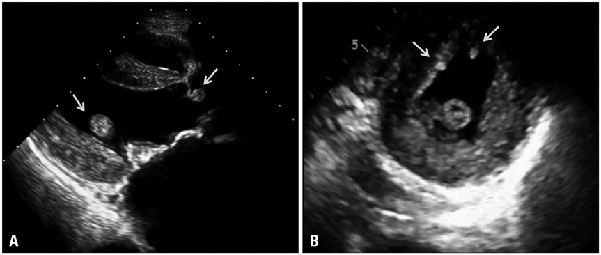
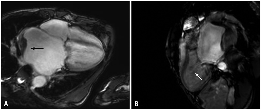
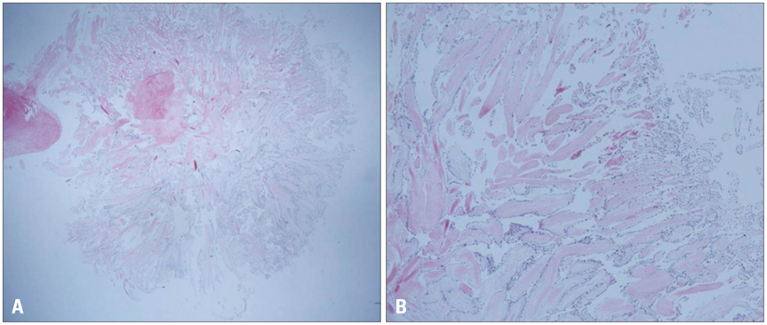
- Cardiac papillary fibroelastomas (CPF) are benign cardiac tumors and usually discovered incidentally during echocardiography. This report describes the case of a 68-year-old man, referred to cardiology for multiple masses of the left ventricle and left atrium. The transthoracic echocardiography revealed multiple oscillating masses in the left ventricle and aortic valve, non-mobile mass in the left atrium with severe mitral stenosis and moderate aortic regurgitation. The patient underwent surgical resection of the masses with valve replacements. Histopathologic examination confirmed the diagnosis of CPF in the left ventricle and aortic valve, thrombus in the left atrium.
Keyword
MeSH Terms
Figure
Reference
-
1. Edwards FH, Hale D, Cohen A, Thompson L, Pezzella AT, Virmani R. Primary cardiac valve tumors. Ann Thorac Surg. 1991; 52:1127–1131.
Article2. Klarich KW, Enriquez-Sarano M, Gura GM, Edwards WD, Tajik AJ, Seward JB. Papillary fibroelastoma: echocardiographic characteristics for diagnosis and pathologic correlation. J Am Coll Cardiol. 1997; 30:784–790.
Article3. Mutlu H, Demir IE, Leppo J, Levy WK. Nonsurgical management of a left ventricular pedunculated papillary fibroelastoma: a case report. J Am Soc Echocardiogr. 2008; 21:877.e4–877.e7.
Article4. Seol SH, Kim DS, Han YC, Kim KH, Kim YB, Kim DK, Ung-Kim , Yang TH, Kim DK, Kim DI. Nonsurgical management of a tricuspid valvular pedunculated papillary fibroelastoma. Cardiovasc Ultrasound. 2009; 7:44.
Article5. Gowda RM, Khan IA, Nair CK, Mehta NJ, Vasavada BC, Sacchi TJ. Cardiac papillary fibroelastoma: a comprehensive analysis of 725 cases. Am Heart J. 2003; 146:404–410.
Article6. Samoun M, Sansone F, Burlo M, Calafiore AM. Papillary fibroelastoma of the anterolateral papillary muscle: an unusual case. J Cardiovasc Med (Hagerstown). 2006; 7:830–832.
Article7. Park JH, Seol SH, Cho HJ, Park SH, Kim DK, Kim U, Yang TH, Kim DK, Kim DI, Kim DS. Papillary fibroelastoma presenting as a left ventricular mass. J Cardiovasc Ultrasound. 2010; 18:66–69.
Article8. Pomerance A. Papillary "tumours" of the heart valves. J Pathol Bacteriol. 1961; 81:135–140.
Article9. Saloura V, Grivas PD, Sarwar AB, Gorodin P, Ledley GS. Papillary fibroelastomas: innocent bystanders or ignored culprits? Postgrad Med. 2009; 121:131–138.
Article10. Grebenc ML, Rosado de Christenson ML, Burke AP, Green CE, Galvin JR. Primary cardiac and pericardial neoplasms: radiologic-pathologic correlation. Radiographics. 2000; 20:1073–1103. quiz 1110-1, 1112.
Article11. Fishbein MC, Ferrans VJ, Roberts WC. Endocardial papillary elastofibromas. Histologic, histochemical, and electron microscopical findings. Arch Pathol. 1975; 99:335–341.
- Full Text Links
- Actions
-
Cited
- CITED
-
- Close
- Share
- Similar articles
-
- Multiple Cardiac Papillary Fibroelastoma of the Aortic Valve
- Early surgical intervention for unusually located cardiac fibroelastomas
- A Case of Multiple Systemic Embolism Associatied with Left Atrial Free-Floating Ball Thrombus
- Acute Ischemic Stroke Caused by Detachment of Cardiac Papillary Fibroelastomas
- Papillary Fibroelastoma of Pulmonary Valve with Congestive Heart Failure: A case report


