Early surgical intervention for unusually located cardiac fibroelastomas
- Affiliations
-
- 1Department of Thoracic and Cardiovascular Surgery, Inje University Sanggye Paik Hospital, Inje University School of Medicine, Seoul, Korea
- 2Division of Cardiology, Department of Internal Medicine, Inje University Sanggye Paik Hospital, Inje University School of Medicine, Seoul, Korea
- 3Department of Pathology, Inje University Sanggye Paik Hospital, Inje University School of Medicine, Seoul, Korea
- KMID: 2508183
- DOI: http://doi.org/10.12701/yujm.2020.00556
Abstract
- Papillary fibroelastomas are the second most common primary cardiac tumor in adults. Over 80% of fibroelastomas occur on the cardiac valves, usually on the left side of the heart, while the remaining lesions are typically scattered throughout the atria and ventricles. Although the optimal timing for surgery is controversial and depends on tumor size and location, prompt surgical resection is warranted in patients at high risk of embolism. A tumor on the cardiac valve can be removed using the slicing excision technique without leaflet injury. Here we present two cases of papillary fibroelastomas occurring on the ventricular surface of the aortic valve and in the right ventricle.
Keyword
Figure
-
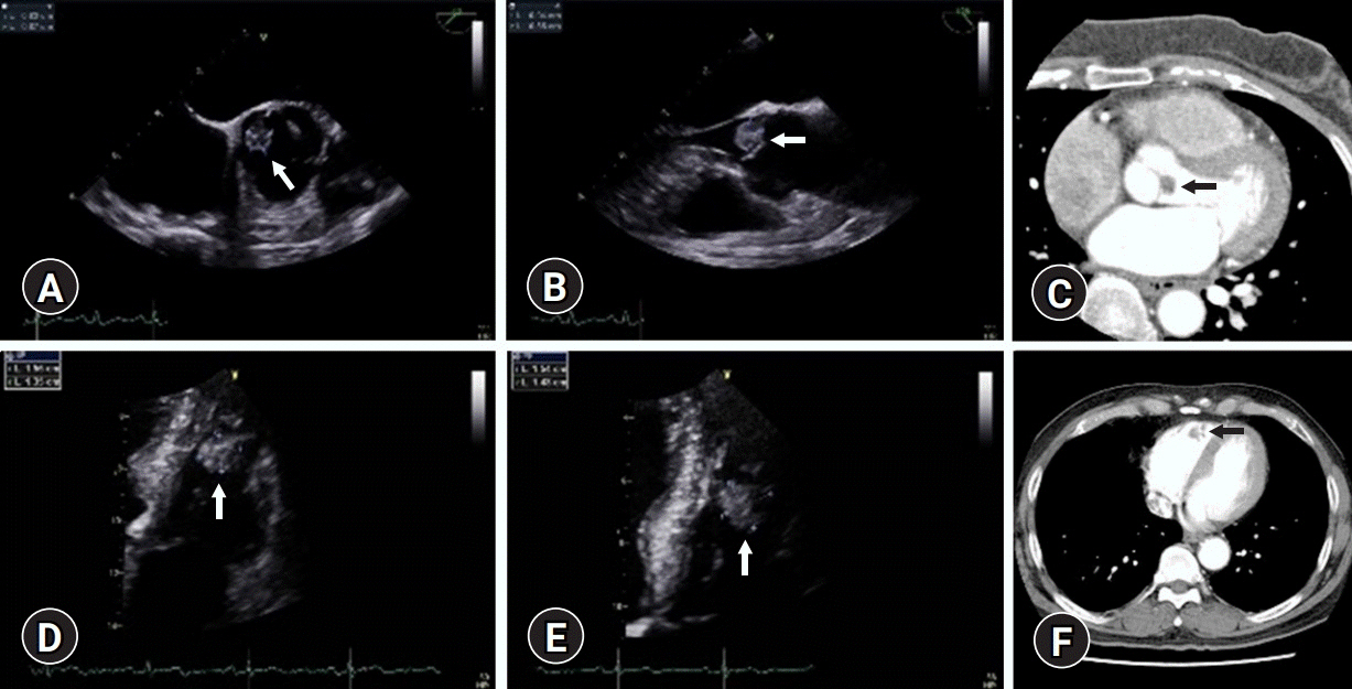
Fig. 1. Radiological findings of case 1 (A–C) and case 2 (D–F). (A, B) Preoperative echocardiograhy shows a mobile mass (arrow) on the ventricular surface of the noncoronary cusp of the aortic valve. (C) Computed tomography image shows a mass lesion (arrow) on the aortic valve. (D, E) Preoperative echocardiography shows a round mass (arrow) in the right ventricle. (F) Computed tomography shows a mass lesion in the right ventricle (arrow).
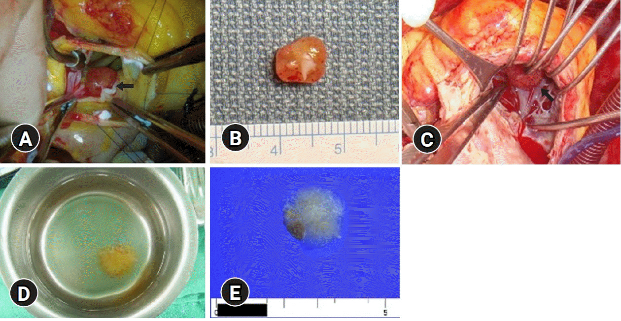
Fig. 2. Operative and gross findings of case 1 (A, B) and case 2 (C–E). (A) The mass is morphologically round and attached to the ventricular surface of the noncoronary cusp of the aortic valve (arrow). (B) A pinkish round mass measures 0.9 cm in the largest dimension. One tip shows a piece of whitish fibrous tissue that might be attached to the aortic valve. (C) The mass is morphologically round and attached to the right ventricular wall (arrow). (D) Immersion in water after resection reveals a typical sea anemone appearance. (E) A whitish papillary mass measures 1.5 cm in the largest dimension. (E) One tip shows a piece of brownish muscular tissue that might be attached to the myocardial wall.
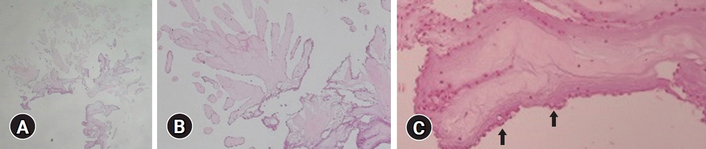
Fig. 3. Histopathological findings. (A) A papillary mass at low power shows numerous arborizing fronds from a common central stalk (hematoxylin and eosin [H&E] stain, ×10). (B) The papillae consist of avascular fronds with hyalinized stroma and partly denuded lining cells (H&E stain, ×40). (C) The fronds are coated by a single endothelial layer (arrows) (H&E stain, ×100).
Reference
-
References
1. Gowda RM, Khan IA, Nair CK, Mehta NJ, Vasavada BC, Sacchi TJ. Cardiac papillary fibroelastoma: a comprehensive analysis of 725 cases. Am Heart J. 2003; 146:404–10.
Article2. Sun JP, Asher CR, Yang XS, Cheng GG, Scalia GM, Massed AG, et al. Clinical and echocardiographic characteristics of papillary fibroelastomas: a retrospective and prospective study in 162 patients. Circulation. 2001; 103:2687–93.
Article3. Grinda JM, Couetil JP, Chauvaud S, D'Attellis N, Berrebi A, Fabiani JN, et al. Cardiac valve papillary fibroelastoma: surgical excision for revealed or potential embolization. J Thorac Cardiovasc Surg. 1999; 117:106–10.
Article4. Ngaage DL, Mullany CJ, Daly RC, Dearani JA, Edwards WD, Tazelaar HD, et al. Surgical treatment of cardiac papillary fibroelastoma: a single center experience with eighty-eight patients. Ann Thorac Surg. 2005; 80:1712–8.
Article5. Tamin SS, Maleszewski JJ, Scott CG, Khan SK, Edwards WD, Bruce CJ, et al. Prognostic and bioepidemiologic implications of papillary fibroelastomas. J Am Coll Cardiol. 2015; 65:2420–9.
Article6. Díaz-Antón B, González Pinto Á, Parra Jiménez FJ, Cuerpo Caballero G, Pérez Rodríguez F, Solís Martín J. Recurrent cardiac fibroelastoma. Is it really a benign tumor? Rev Esp Cardiol (Engl Ed). 2018; 71:685–7.
Article
- Full Text Links
-
- Actions
-
Cited
- CITED
-
- Close
- Share
-
- Similar articles
-
- Multiple Cardiac Papillary Fibroelastoma of the Aortic Valve
- Acute Ischemic Stroke Caused by Detachment of Cardiac Papillary Fibroelastomas
- Multiple Papillary Fibroelastomas and Thrombus in the Left Heart
- Early Detection and Intervention of Autism Spectrum Disorder
- Recurrent Cardiac Tamponade Complicated by Coronary Intervention
