J Cardiovasc Ultrasound.
2012 Jun;20(2):90-96. 10.4250/jcu.2012.20.2.90.
Impact of Arterial Stiffness on Regional Myocardial Function Assessed by Speckle Tracking Echocardiography in Patients with Hypertension
- Affiliations
-
- 1Department of Cardiology, Ulsan University Hospital, Ulsan, Korea.
- 2Department of Cardiology, University of Ulsan College of Medicine, Asan Medical Center, Seoul, Korea. sjkang@amc.seoul.kr
- 3Department of Cardiology, Ajou University Medical Center, Suwon, Korea.
- KMID: 2177372
- DOI: http://doi.org/10.4250/jcu.2012.20.2.90
Abstract
- BACKGROUND
Arterial stiffening may affect regional myocardial function in hypertensive patients with normal ejection fraction (EF).
METHODS
Brachial-ankle pulse wave velocity (PWV) was measured in 70 patients, of mean age 48 +/- 14 years, with untreated hypertension and EF > 55%. Using two-dimensional-speckle tracking echocardiography, we measured longitudinal and circumferential strain (epsilon) and strain rate (SR). Basal and apical rotations were measured using short axis views.
RESULTS
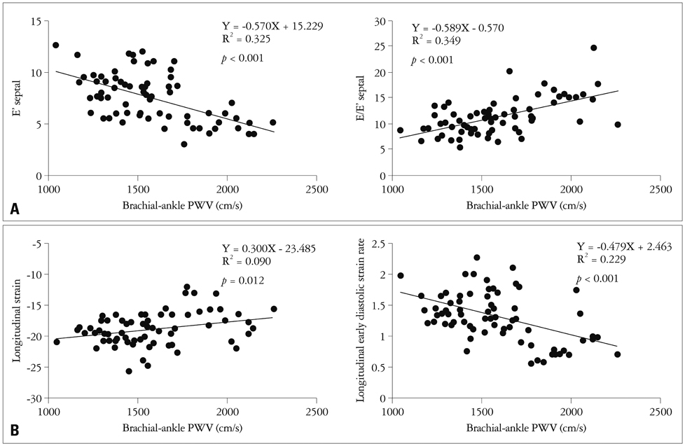
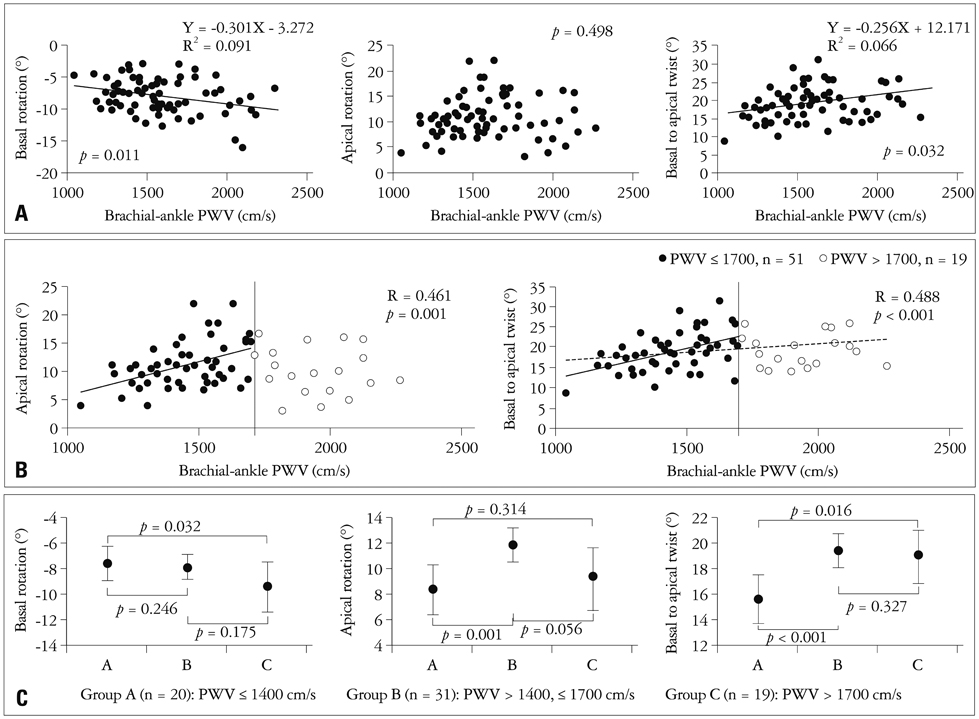
The mean systolic and diastolic blood pressure in these patients was 152 +/- 15 mmHg and 92 +/- 11 mmHg, respectively. The mean value of PWV was 1578 +/- 274 cm/s. PWV significantly correlated with age (r = 0.682, p < 0.001), body mass index (r = -0.330, p = 0.005), systolic blood pressure (r = 0.386, p = 0.001) and pulse pressure (r = 0.509, p < 0.001). PWV also significantly correlated with septal E' velocity (r = -0.570, p < 0.001), E/A ratio (r = -0.414, p < 0.001), E/E' ratio (r = 0.589, p < 0.001), systolic global longitudinal epsilon (r = 0.300, p = 0.012) and early diastolic SR (SRE) (r = -0.479, p < 0.001) suggesting impaired abnormal relaxation. PWV was also correlated with basal rotation (r = -0.301, p = 0.011) and basal-to-apical twist (r = -0.256, p = 0.032). The increases in apical rotation and basal-to-apical twist were attenuated in patients with PWV > 1700 cm/s compared to those with PWV < or = 1400 cm/s or those with PWV 1400-1700 cm/s.
CONCLUSION
In hypertensive patients with normal ejection fraction, arterial stiffening contributes to impaired systolic and diastolic function of the regional myocardium. Compensatory increases in ventricular twist were diminished in patients with advanced stage of vascular stiffening.
MeSH Terms
Figure
Reference
-
1. Moore JE Jr, Xu C, Glagov S, Zarins CK, Ku DN. Fluid wall shear stress measurements in a model of the human abdominal aorta: oscillatory behavior and relationship to atherosclerosis. Atherosclerosis. 1994. 110:225–240.
Article2. Chen CH, Nakayama M, Nevo E, Fetics BJ, Maughan WL, Kass DA. Coupled systolic-ventricular and vascular stiffening with age: implications for pressure regulation and cardiac reserve in the elderly. J Am Coll Cardiol. 1998. 32:1221–1227.3. Kass DA. Ventricular arterial stiffening: integrating the pathophysiology. Hypertension. 2005. 46:185–193.4. Kawaguchi M, Hay I, Fetics B, Kass DA. Combined ventricular systolic and arterial stiffening in patients with heart failure and preserved ejection fraction: implications for systolic and diastolic reserve limitations. Circulation. 2003. 107:714–720.
Article5. Leitman M, Lysyansky P, Sidenko S, Shir V, Peleg E, Binenbaum M, Kaluski E, Krakover R, Vered Z. Two-dimensional strain-a novel software for real-time quantitative echocardiographic assessment of myocardial function. J Am Soc Echocardiogr. 2004. 17:1021–1029.
Article6. Serri K, Reant P, Lafitte M, Berhouet M, Le Bouffos V, Roudaut R, Lafitte S. Global and regional myocardial function quantification by two-dimensional strain: application in hypertrophic cardiomyopathy. J Am Coll Cardiol. 2006. 47:1175–1181.
Article7. Lang RM, Bierig M, Devereux RB, Flachskampf FA, Foster E, Pellikka PA, Picard MH, Roman MJ, Seward J, Shanewise JS, Solomon SD, Spencer KT, Sutton MS, Stewart WJ. Chamber Quantification Writing Group. American Society of Echocardiography's Guidelines and Standards Committee. European Association of Echocardiography. Recommendations for chamber quantification: a report from the American Society of Echocardiography's Guidelines and Standards Committee and the Chamber Quantification Writing Group, developed in conjunction with the European Association of Echocardiography, a branch of the European Society of Cardiology. J Am Soc Echocardiogr. 2005. 18:1440–1463.
Article8. Nagueh SF, Appleton CP, Gillebert TC, Marino PN, Oh JK, Smiseth OA, Waggoner AD, Flachskampf FA, Pellikka PA, Evangelista A. Recommendations for the evaluation of left ventricular diastolic function by echocardiography. J Am Soc Echocardiogr. 2009. 22:107–133.
Article9. Frenneaux M, Williams L. Ventricular-arterial and ventricular-ventricular interactions and their relevance to diastolic filling. Prog Cardiovasc Dis. 2007. 49:252–262.
Article10. Saeki A, Recchia F, Kass DA. Systolic flow augmentation in hearts ejecting into a model of stiff aging vasculature. Influence on myocardial perfusion-demand balance. Circ Res. 1995. 76:132–141.
Article11. Kelly RP, Tunin R, Kass DA. Effect of reduced aortic compliance on cardiac efficiency and contractile function of in situ canine left ventricle. Circ Res. 1992. 71:490–502.
Article12. Lartaud-Idjouadiene I, Lompré AM, Kieffer P, Colas T, Atkinson J. Cardiac consequences of prolonged exposure to an isolated increase in aortic stiffness. Hypertension. 1999. 34:63–69.
Article13. Kang SJ, Lim HS, Choi BJ, Choi SY, Hwang GS, Yoon MH, Tahk SJ, Shin JH. Longitudinal strain and torsion assessed by two-dimensional speckle tracking correlate with the serum level of tissue inhibitor of matrix metalloproteinase-1, a marker of myocardial fibrosis, in patients with hypertension. J Am Soc Echocardiogr. 2008. 21:907–911.
Article14. Notomi Y, Lysyansky P, Setser RM, Shiota T, Popović ZB, Martin-Miklovic MG, Weaver JA, Oryszak SJ, Greenberg NL, White RD, Thomas JD. Measurement of ventricular torsion by two-dimensional ultrasound speckle tracking imaging. J Am Coll Cardiol. 2005. 45:2034–2041.
Article15. Nagel E, Stuber M, Burkhard B, Fischer SE, Scheidegger MB, Boesiger P, Hess OM. Cardiac rotation and relaxation in patients with aortic valve stenosis. Eur Heart J. 2000. 21:582–589.
Article16. Sandstede JJ, Johnson T, Harre K, Beer M, Hofmann S, Pabst T, Kenn W, Voelker W, Neubauer S, Hahn D. Cardiac systolic rotation and contraction before and after valve replacement for aortic stenosis: a myocardial tagging study using MR imaging. AJR Am J Roentgenol. 2002. 178:953–958.
Article17. Chung J, Abraszewski P, Yu X, Liu W, Krainik AJ, Ashford M, Caruthers SD, McGill JB, Wickline SA. Paradoxical increase in ventricular torsion and systolic torsion rate in type I diabetic patients under tight glycemic control. J Am Coll Cardiol. 2006. 47:384–390.
Article18. Götte MJ, Germans T, Rüssel IK, Zwanenburg JJ, Marcus JT, van Rossum AC, van Veldhuisen DJ. Myocardial strain and torsion quantified by cardiovascular magnetic resonance tissue tagging: studies in normal and impaired left ventricular function. J Am Coll Cardiol. 2006. 48:2002–2011.
Article19. Kim HK, Sohn DW, Lee SE, Choi SY, Park JS, Kim YJ, Oh BH, Park YB, Choi YS. Assessment of left ventricular rotation and torsion with two-dimensional speckle tracking echocardiography. J Am Soc Echocardiogr. 2007. 20:45–53.
Article
- Full Text Links
- Actions
-
Cited
- CITED
-
- Close
- Share
- Similar articles
-
- Current Status of 3-Dimensional Speckle Tracking Echocardiography: A Review from Our Experiences
- Non-invasive assessment of vascular alteration using ultrasound
- Differences in left ventricular functional adaptation to arterial stiffness and neurohormonal activation in patients with hypertension: a study with two-dimensional layer-specific speckle tracking echocardiography
- Global and Regional Right Ventricular Function Investigation by 2D Speckle Tracking Echocardiography in Congenital Heart Disease Patients With Pulmonary Arterial Hypertension
- Global longitudinal strain manually measured from mid‑myocardial lengths is a reliable alternative to speckle tracking global longitudinal strain


