Cases of Hemolytic Anemia with Periprosthetic Leaks Evaluated by Real-Time 3-Dimensional Transesophageal Echocardiography
- Affiliations
-
- 1Department of Cardiovascular Medicine, Konkuk University Medical Center, Seoul, Korea. yang.hyun@kuh.ac.kr
- 2Department of Anesthesiology, Konkuk University Medical Center, Seoul, Korea.
- 3Department of Internal Medicine, Pusan National University College of Medicine, Busan, Korea.
- 4Department of Cardiothoracic Surgery, Pusan National University College of Medicine, Busan, Korea.
- KMID: 2177342
- DOI: http://doi.org/10.4250/jcu.2012.20.1.52
Abstract
- Hemolytic anemia is recognized as a rare complication of mitral valve replacement or repair. We report on a 44-year-old man with shortness of breath and hemolytic anemia, 23 years after mitral valve replacement (Hall-Kaster), and a 63-year-old woman diagnosed of hemolytic anemia, 4 years after mitral and tricuspid annuloplasty (Tailor ring, An-core ring). Routine 2-dimensional transthoracic echocardiography revealed paravalvular leakage around the prosthesis. Subsequent real-time 3-dimensional (3D)transesophageal echocardiography helped the perceptional appreciation of the leakage and the measuring of the regurgitant orifice area using the anatomically correct plane. Surgical findings of each case fit those of 3D volumetric images.
MeSH Terms
Figure
-

Fig. 1 A: A real-time 3-dimensional (3D) transesophageal echocardiography shows a paravalvular leak (arrows) outside of the mechanical mitral prosthesis (mid-posterior) in en-face view from a left atrial perspective, obtained in a single heart beat using 3D zoom mode. B: A full-volume 3D color volume reveals a significant systolic paravalvular regurgitant jet (arrows) and small physiological valvular regurgitant jets (arrowheads), acquired in 7 electrocardiography-gated cardiac cycles.
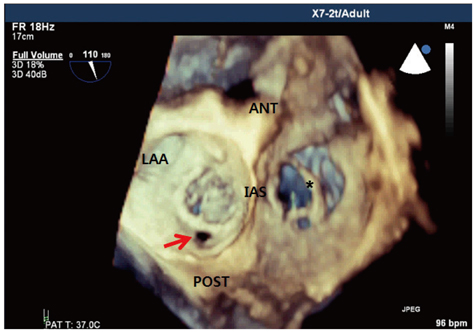
Fig. 2 This one 3-dimensional full-volume image shows the mitral posterior ring and peri-ring tissue defect (arrow) and the tricuspid annuloplasty ring prosthesis (asterisk) which was more than half detached from the anatomical annulus. ANT: anterior, POST: posterior, LAA: left atrial appendage, IAS: interatrial septum.
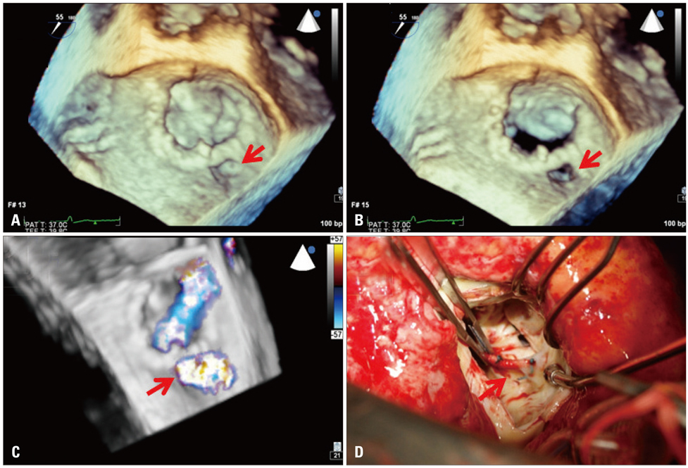
Fig. 3 A real-time 3-dimensional (3D) transesophageal echocardiography zoom image demonstrates a peri-ring tissue defect (arrow) of the mitral posterior ring (arrow) on surgeon's view in end-systole (A) and mid-diastole (B). A full-volume 3D color image reveals the regurgitant jet from the peri-ring defect (arrow), differentiated from valvular regurgitation (C); this view is similar to what the surgeon saw (D).
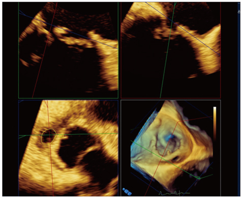
Fig. 4 Postprocessing of the live 3-dimensional transesophageal echocardiography volumetric image using the multi-planar rendering (MPR) mode in QLAB software (Philips Medical Systems, Andover, MA, USA) measures the peri-ring regurgitation orifice area using the anatomically correct plane as 0.61 cm2.
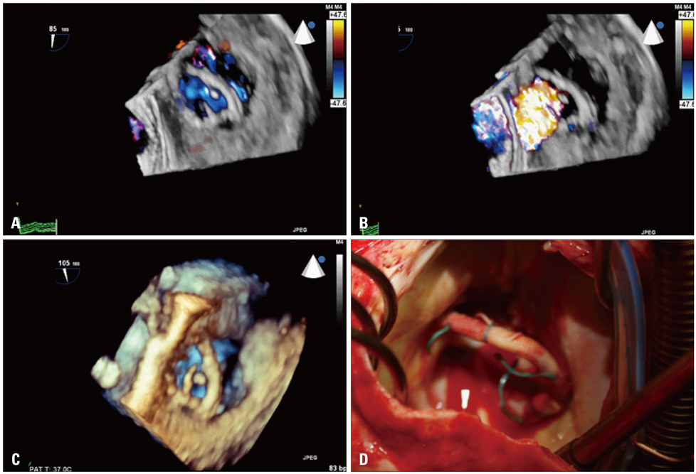
Fig. 5 A full-volume 3-dimensional (3D) color transesophageal echocardiography (TEE) image shows the tricuspid ring prosthesis in diastole (A), combined with regurgitation through the ring prosthesis during systole (B). A real-time 3D TEE zoom image reveals the tricuspid ring prosthesis and several thread-like strands caused by detachment from the native annulus (C), which revelation is confirmed in the surgical findings (D).
Reference
-
1. Edmunds LH Jr, Clark RE, Cohn LH, Grunkemeier GL, Miller DC, Weisel RD. Guidelines for reporting morbidity and mortality after cardiac valvular operations. Ad Hoc Liaison Committee for Standardizing Definitions of Prosthetic Heart Valve Morbidity of The American Association for Thoracic Surgery and The Society of Thoracic Surgeons. J Thorac Cardiovasc Surg. 1996. 112:708–711.
Article2. Maraj R, Jacobs LE, Ioli A, Kotler MN. Evaluation of hemolysis in patients with prosthetic heart valves. Clin Cardiol. 1998. 21:387–392.
Article3. Garcia MJ, Vandervoort P, Stewart WJ, Lytle BW, Cosgrove DM 3rd, Thomas JD, Griffin BP. Mechanisms of hemolysis with mitral prosthetic regurgitation. Study using transesophageal echocardiography and fluid dynamic simulation. J Am Coll Cardiol. 1996. 27:399–406.
Article4. Iguro Y, Moriyama Y, Yamaoka A, Yamashita M, Shimokawa S, Toyohira H, Taira A. Clinical experience of 473 patients with the omnicarbon prosthetic heart valve. J Heart Valve Dis. 1999. 8:674–679.5. Skoularigis J, Essop MR, Skudicky D, Middlemost SJ, Sareli P. Frequency and severity of intravascular hemolysis after left-sided cardiac valve replacement with Medtronic Hall and St. Jude Medical prostheses, and influence of prosthetic type, position, size and number. Am J Cardiol. 1993. 71:587–591.
Article6. Mecozzi G, Milano AD, De Carlo M, Sorrentino F, Pratali S, Nardi C, Bortolotti U. Intravascular hemolysis in patients with new-generation prosthetic heart valves: a prospective study. J Thorac Cardiovasc Surg. 2002. 123:550–556.
Article7. Demirsoy E, Yilmaz O, Sirin G, Baran T, Tekin S, Sener D, Sonmez B. Hemolysis after mitral valve repair: a report of five cases and literature review. J Heart Valve Dis. 2008. 17:24–30.8. Yang HS, Bansal RC, Mookadam F, Khandheria BK, Tajik AJ, Chandrasekaran K. American Society of Echocardiography. Practical guide for three-dimensional transthoracic echocardiography using a fully sampled matrix array transducer. J Am Soc Echocardiogr. 2008. 21:979–989. quiz 1081-2.
Article9. Sugeng L, Shernan SK, Weinert L, Shook D, Raman J, Jeevanandam V, DuPont F, Fox J, Mor-Avi V, Lang RM. Real-time three-dimensional transesophageal echocardiography in valve disease: comparison with surgical findings and evaluation of prosthetic valves. J Am Soc Echocardiogr. 2008. 21:1347–1354.
Article10. Yang HS, Srivathsan K, Wissner E, Chandrasekaran K. Images in cardiovascular medicine. Real-time 3-dimensional transesophageal echocardiography: novel utility in atrial fibrillation ablation with a prosthetic mitral valve. Circulation. 2008. 117:e304–e305.
- Full Text Links
-
- Actions
-
Cited
- CITED
-
- Close
- Share
-
- Similar articles
-
- Horseshoe-like Shaped Atrial Septal Defects Confirmed on Three-Dimensional Transesophageal Echocardiography
- Role of modern 3D echocardiography in valvular heart disease
- Real-time Three Dimensional Echocardiography Clinical Implications and The Perspective
- Usefulness of intraoperative real-time three-dimensional transesophageal echocardiography in preprocedural evaluation of cortriatriatum: a case report
- Advances in the Evaluation of Mitral Complex Geometry:Insights from Transthoracic Real-time Three-dimensional Echocardiography
