Development of implant loading device for animal study about various loading protocol: a pilot study
- Affiliations
-
- 1Department of Prosthodontics, Yonsei University College of Dentistry, Seoul, Korea. jfshim@yuhs.ac
- 2Department of Periodontology, Yonsei University College of Dentistry, Seoul, Korea.
- KMID: 2176422
- DOI: http://doi.org/10.4047/jap.2012.4.4.227
Abstract
- PURPOSE
The aims of this pilot study were to introduce implant loading devices designed for animal study and to evaluate the validity of the load transmission ability of the loading devices.
MATERIALS AND METHODS
Implant loading devices were specially designed and fabricated with two implant abutments and cast metal bars, and orthodontic expansion screw. In six Beagles, all premolars were extracted and two implants were placed in each side of the mandibles. The loading device was inserted two weeks after the implant placement. According to the loading protocol, the load was applied to the implants with different time and method,simulating early, progressive, and delayed loading. The implants were clinically evaluated and the loading devices were removed and replaced to the master cast, followed by stress-strain analysis. Descriptive statistics of remained strain (microepsilon) was evaluated after repeating three cycles of the loading device activation. Statistic analysis was performed using nonparametric, independent t-test with 5% significance level and Friedman's test was also used for verification.
RESULTS
The loading devices were in good action. However, four implants in three Beagles showed loss of osseointegration. In stress-strain analysis, loading devices showed similar amount of increase in the remained strain after applying 1-unit load for three times.
CONCLUSION
Specialized design of the implant loading device was introduced. The loading device applied similar amount of loads near the implant after each 1-unit loading. However, the direction of the loads was not parallel to the long axis of the implants as predicted before the study.
Keyword
MeSH Terms
Figure
-
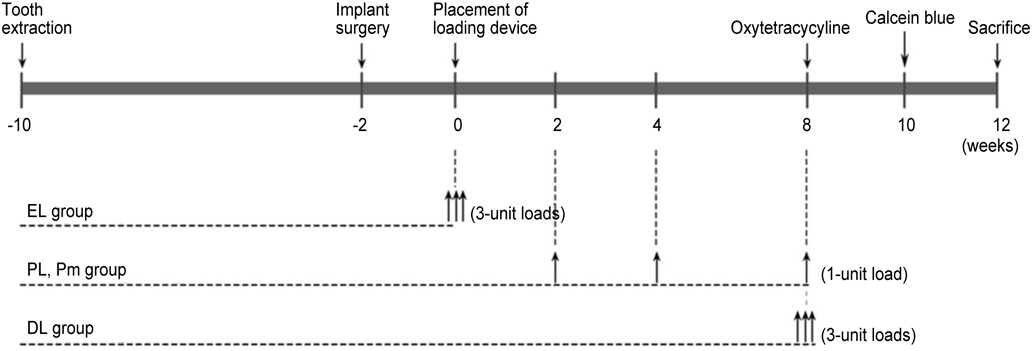
Fig. 1 Loading protocols. EL: early loading, PL: periodic loading, Pm: periodic loading, rotation mobile, DL: delayed loading.

Fig. 2 Implant placement and impression taking. A: The implants were placed 10 mm apart, remaining 2 mm of buccal and lingual cortex, B: Impression was taken with impression copings and pattern resin.

Fig. 3 Diagram of the loading device. A: A schematic illustration of the loading device, B: The direction of force applied to the implant and the surrounding alveolar bone.
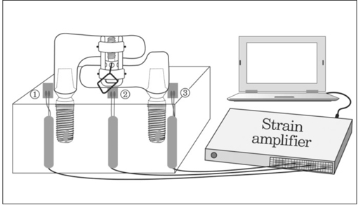
Fig. 4 Measurement of efficiency of the loading device with strain gauge. Three strain gauges were attached on the distal side of both implants and on the center of both implants.
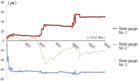
Fig. 5 Representative illustration of stress-strain curve over time.
Reference
-
1. Testori T, Wiseman L, Woolfe S, Porter SS. A prospective multicenter clinical study of the Osseotite implant: four-year interim report. Int J Oral Maxillofac Implants. 2001. 16:193–200.2. Cochran DL, Morton D, Weber HP. Consensus statements and recommended clinical procedures regarding loading protocols for endosseous dental implants. Int J Oral Maxillofac Implants. 2004. 19:109–113.3. Ledermann P. Bar-prosthetic management of the edentulous mandible by means of plasma-coated implantation with titanium screws. Dtsch Zahnarztl Z. 1979. 34:907–911.4. Romanos G, Froum S, Hery C, Cho SC, Tarnow D. Survival rate of immediately vs delayed loaded implants: analysis of the current literature. J Oral Implantol. 2010. 36:315–324.5. Rocci A, Martignoni M, Burgos PM, Gottlow J, Sennerby L. Histology of retrieved immediately and early loaded oxidized implants: light microscopic observations after 5 to 9 months of loading in the posterior mandible. Clin Implant Dent Relat Res. 2003. 5:88–98.6. Romanos GE, Nentwig GH. Immediate versus delayed functional loading of implants in the posterior mandible: a 2-year prospective clinical study of 12 consecutive cases. Int J Periodontics Restorative Dent. 2006. 26:459–469.7. Hoshaw SJ, Brunski JB, Cochran GVB. Mechanical loading of Branemark implants affects interfacial bone modeling and remodeling. Int J Oral Maxillofac Implants. 1994. 9:345–360.8. Frost HM. Wolff's Law and bone's structural adaptations to mechanical usage: an overview for clinicians. Angle Orthod. 1994. 64:175–188.9. Binon PP, Sullivan DY. Provisional fixed restorations technique for osseointegrated implants. J Calif Dent Assoc. 1990. 18:23–30.10. Misch CE. Contemporary Implant Dentistry. 1999. 2nd ed. St Louis: Mosby;595–608.11. Appleton RS, Nummikoski PV, Pigno MA, Cronin RJ, Chung KH. A radiographic assessment of progressive loading on bone around single osseointegrated implants in the posterior maxilla. Clin Oral Implants Res. 2005. 16:161–167.12. Roberts WE. Bone tissue interface. J Dent Educ. 1988. 52:804–809.13. Jayme SJ, de Oliveira RR, Muglia VA, Novaes AB Jr, Ribeiro RF. The effects of different loading times on the bone response around dental implants: a histomorphometric study in dogs. Int J Oral Maxillofac Implants. 2010. 25:473–481.14. Gostović AS, Todorović A, Lazić V, Todorović A, Milinković I, Leković V. Immediate implant loading with fixed dental restorations-an animal model study. Vojnosanit Pregl. 2012. 69:181–189.15. Rismanchian M, Attar BM, Razavi SM, Shamsabad AN, Rezaei M. Dental implants immediate loading versus the standard 2-staged protocol: an experimental study in dogs. J Oral Implantol. 2012. 38:3–10.16. Rismanchian M, Bajoghli F, Gholamreza T, Razavi M. Clinical, histological and histomorphometrical evaluation of early loaded implants (an animal study). J Oral Implantol. 2012. 01. 03.17. Ivanoff CJ, Sennerby L, Lekholm U. Influence of initial implant mobility on the integration of titanium implants. An experimental study in rabbits. Clin Oral Implants Res. 1996. 7:120–127.18. Tanck E, Homminga J, van Lenthe GH, Huiskes R. Increase in bone volume fraction precedes architectural adaptation in growing bone. Bone. 2001. 28:650–654.19. Turner CH. Three rules for bone adaptation to mechanical stimuli. Bone. 1998. 23:399–407.20. Gotfredsen K, Berglundh T, Lindhe J. Bone reactions adjacent to titanium implants subjected to static load. A study in the dog (I). Clin Oral Implants Res. 2001. 12:1–8.21. Brunski JB. Biomechanical factors affecting the bone-dental implant interface. Clin Mater. 1992. 10:153–201.22. Szmukler-Moncler S, Piattelli A, Favero GA, Dubruille JH. Considerations preliminary to the application of early and immediate loading protocols in dental implantology. Clin Oral Implants Res. 2000. 11:12–25.23. Mishra M, Ozawa S, Masuda T, Yoshioka F, Tanaka Y. Finite element study on the effect of abutment length and material on implant bone interface against dynamic loading. J Adv Prosthodont. 2011. 3:140–144.24. Sohn BS, Heo SJ, Koak JY, Kim SK, Lee SY. Strain of implants depending on occlusion types in mandibular implant-supported fixed prostheses. J Adv Prosthodont. 2011. 3:1–9.25. Jemt T, Book K. Prosthesis misfit and marginal bone loss in edentulous implant patients. Int J Oral Maxillofac Implants. 1996. 11:620–625.26. Duyck J, Rønold HJ, Van Oosterwyck H, Naert I, Vander Sloten J, Ellingsen JE. The influence of static and dynamic loading on marginal bone reactions around osseointegrated implants: an animal experimental study. Clin Oral Implants Res. 2001. 12:207–218.
- Full Text Links
-
- Actions
-
Cited
- CITED
-
- Close
- Share
-
- Similar articles
-
- Effect of cyclic loading and retightening on reverse torque value in external and internal implants
- Recent advances in dental implants
- The effect of loading time on the stability of mini-implant
- Pull-off resistance of a screwless implantabutment connection and surface evaluation after cyclic loading
- The influence of abutment screw tightening timing and DLC coating of conical connection implant system
