Quantitative comparison of permeability in the adhesive interface of four adhesive systems
- Affiliations
-
- 1Department of Conservative Dentistry and Dental Research Institute, School of Dentistry, Seoul National University, Korea. hhson@snu.ac.kr
- 2Geochronology Team, Korea Basic Science Institute, Korea.
- 3Craniomaxillofacial Life Science(BK 21), School of Dentistry, Seoul National University, Korea.
- KMID: 2176043
- DOI: http://doi.org/10.5395/JKACD.2009.34.1.051
Abstract
- The purpose of this study was to perform quantitative comparisons of water permeable zones in both the adhesive and the hybrid layer before and after thermocycling in order to assess the integrity of the bonding interface. Twenty eight flat dentin surfaces were bonded with a light-cured composite resin using one of four commercial adhesives [OptiBond FL (OP), AdheSE (AD), Clearfil SE Bond (CL), and Xeno III (XE)]. These were sectioned into halves and subsequently cut to yield 2-mm thick specimens; one specimen for control and the other subjected to thermocycling for 10,000 cycles. After specimens were immersed in ammoniacal silver nitrate for 24 h and exposed to a photo developing solution for 8 h, the bonded interface was analyzed by scanning electron microscopy (SEM) and wavelength dispersive spectrometry (WDS) at five locations per specimen. Immediately after bonding, the adhesive layer of OP showed the lowest silver uptake, followed by CL, AD, and XE in ascending order (p < 0.0001); the hybrid layer of CL had the lowest silver content among the groups (p = 0.0039). After thermocycling, none of the adhesives manifested a significant increase of silver in either the adhesive or the hybrid layer. SEM demonstrated the characteristic silver penetrated patterns within the interface. It was observed that integrity of bonding was well maintained in OP and CL throughout the thermocycling process. Adhesive-tooth interfaces are vulnerable to hydrolytic degradation and its permeability varies in different adhesive systems, which may be clinically related to the restoration longevity.
Keyword
MeSH Terms
Figure
-
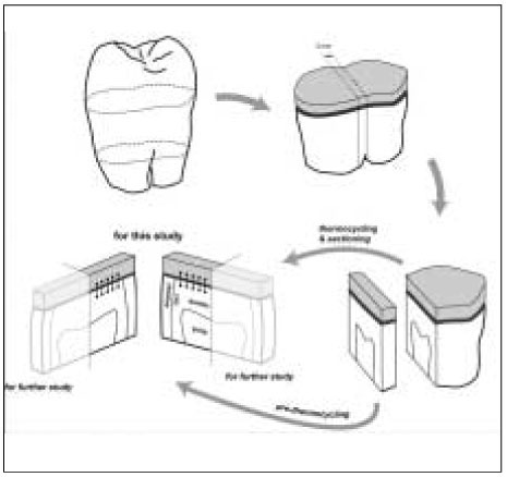
Figure 1 Diagram for specimen preparation.
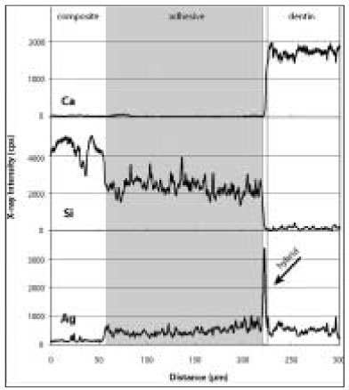
Figure 2 a Elemental analysis of silver (Ag), calcium (Ca), and silica (Si) on an immediate bonding specimen in OptiBond FL group. Note the sharp peak of Ag as demonstrated in the hybrid layer (arrow) where the Ca value started to rise as well.

Figure 2 b An immediate bonding specimen in the Clearfil SE Bond group. Note the increase of Ag in the adhesive layer. The Si level of the adhesive is lower than that in OP because of their different filler composition.
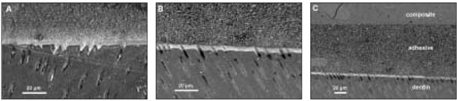
Figure 3 OptiBond FL: (a) SEM micrograph with 1000× magnification of the initial bonding status before thermocycling. Silver uptakes were represented by a dense white image in the hybrid layer and dentinal tubules (b) After thermocycling. SEM showed the adjacent location to where the picture (a) was taken. Silver penetration occurred in a similar pattern. (c) SEM of the same location as (b) with a lower magnification. Integrity of bonding seemed to be maintained between composite resin and dentin throughout thermocycling.
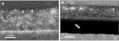
Figure 4 AdheSE: (a) Before thermocycling. Multiple silver droplets were contained within the adhesive layer. Artifact ual crack line from vacuum stress was demonstrated right above the hybrid layer (arrows). (b) The adjacent area aft er thermocycling. Crack propagated further between the adhesive and hybrid layer (white arrow). There was no change in silver accumulation patterns.
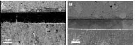
Figure 5 Clearfil SE Bond: (a) Before thermocycling. Silver mostly presented in the thin hybrid layer. (b) The area next to (a) after thermocycling. Bonding interface kept integrated with the silver penetrated hybrid layer in the same manner.

Figure 6 Xeno III [XE] (a) Bonding before thermocycling. It is noticeable that silver penetration occurred in composite/enamel interface as well as in composite/dentin and appeared overall in the adhesive and hybrid layer. (b) Before thermocycling. Dense deposition of silver was spread uniformly along the adhesive and hybrid layer. (c) The adjacent location was shown after thermocycling. Silver particles coalesced in an irregular form.
Reference
-
1. Opdam NJ, Bronkhorst EM, Roeters JM, Loomans BA. A retrospective clinical study on longevity of posterior composite and amalgam restorations. Dent Mater. 2007. 23(1):2–8.
Article2. da Rosa Rodolpho PA, Cenci MS, Donassollo TA, Loguercio AD, Demarco FF. A clinical evaluation of posterior composite restorations: 17-year findings. J Dent. 2006. 34(7):427–435.
Article3. Kramer N, Garcia-Godoy F, Reinelt C, Frankenberger R. Clinical performance of posterior compomer restorations over 4 years. Am J Dent. 2006. 19(1):61–66.4. Manhart J, Chen H, Hamm G, Hickel R. Buonocore Memorial Lecture. Review of the clinical survival of direct and indirect restorations in posterior teeth of the permanent dentition. Oper Dent. 2004. 29(5):481–508.5. De Munck J, Van Landuyt K, Peumans M, Poitevin A, Lambrechts P, Braem M, et al. A critical review of the durability of adhesion to tooth tissue: methods and results. J Dent Res. 2005. 84(2):118–132.
Article6. Tay FR, King NM, Chan KM, Pashley DH. How can nanoleakage occur in self-etching adhesive systems that demineralize and infiltrate simultaneously? J Adhes Dent. 2002. 4(4):255–269.7. De Munck J, Van Landuyt K, Coutinho E, Poitevin A, Peumans M, Lambrechts P, et al. Micro-tensile bond strength of adhesives bonded to Class-I cavity-bottom dentin after thermo-cycling. Dent Mater. 2005. 21(11):999–1007.
Article8. Tulunoglu O, Uctasli MB, Ozdemir S. Coronal microleakage of temporary restorations in previously restored teeth with amalgam and composite. Oper Dent. 2005. 30(3):331–337.9. Hiraishi N, Breschi L, Prati C, Ferrari M, Tagami J, King NM. Technique sensitivity associated with air-drying of HEMA-free, single-bottle, one-step self-etch adhesives. Dent Mater. 2007. 23(4):498–505.
Article10. Reis AF, Bedran-Russo AK, Giannini M, Pereira PN. Interfacial ultramorphology of single-step adhesives: nanoleakage as a function of time. J Oral Rehabil. 2007. 34(3):213–221.
Article11. Reis AF, Giannini M, Pereira PN. Long-term TEM analysis of the nanoleakage patterns in resin-dentin interfaces produced by different bonding strategies. Dent Mater. 2007. 23(9):1164–1172.
Article12. Yuan Y, Shimada Y, Ichinose S, Tagami J. Qualitative analysis of adhesive interface nanoleakage using FESEM/EDS. Dent Mater. 2007. 23(5):561–569.
Article13. Chang J, Platt JA, Yi K, Cochran MA. Quantitative comparison of the water permeable zone among four types of dental adhesives used with a dual-cured composite. Oper Dent. 2006. 31(3):346–353.
Article14. Akimoto N, Yokoyama G, Ohmori K, Suzuki S, Kohno A, Cox CF. Remineralization across the resin-dentin interface: in vivo evaluation with nanoindentation measurements, EDS, and SEM. Quintessence Int. 2001. 32(7):561–570.15. Pashley EL, Agee KA, Pashley DH, Tay FR. Effects of one versus two applications of an unfilled, all-in-one adhesive on dentine bonding. J Dent. 2002. 30:83–90.
Article16. Littell RC. SAS System for Mixed Models. 1996. Cary, NC: SAS Institute Inc;171–227.17. Eckert GJ, Platt JA. A statistical evaluation of microtensile bond strength methodology for dental adhesives. Dent Mater. 2007. 23(3):385–391.
Article18. Reis A, Grande RH, Oliveira GM, Lopes GC, Loguercio AD. A 2-year evaluation of moisture on microtensile bond strength and nanoleakage. Dent Mater. 2007. 23(7):862–870.
Article19. Choi SM, Park SH, Choi KK, Park SJ. Effect of the additional application of a resin layer on dentin bonding using single-step adhesives. J Korean Acad Conserv Dent. 2007. 32:313–326.
Article20. Moszner N, Salz U, Zimmermann J. Chemical aspects of self-etching enamel-dentin adhesives: a systematic review. Dent Mater. 2005. 21(10):895–910.
Article21. Gueders AM, Charpentier JF, Albert AI, Geerts SO. Microleakage after thermocycling of 4 etch and rinse and 3 self-etch adhesives with and without a flowable composite lining. Oper Dent. 2006. 31(4):450–455.
Article22. Inoue S, Koshiro K, Yoshida Y, De Munck J, Nagakane K, Suzuki K, et al. Hydrolytic stability of self-etch adhesives bonded to dentin. J Dent Res. 2005. 84(12):1160–1164.
Article23. Monticelli F, Osorio R, Pisani-Proenca J, Toledano M. Resistance to degradation of resin-dentin bonds using a one-step HEMA-free adhesive. J Dent. 2007. 35(2):181–186.
Article24. Tay FR, Suh BI, Pashley DH, Prati C, Chuang SF, Li F. Factors contributing to the incompatibility between simplified-step adhesives and self-cured or dual-cured composites. Part II. Single-bottle, total-etch adhesive. J Adhes Dent. 2003. 5(2):91–105.25. Cheong C, King NM, Pashley DH, Ferrari M, Toledano M, Tay FR. Incompatibility of self-etch adhesives with chemical/dual-cured composites: two-step vs one-step systems. Oper Dent. 2003. 28(6):747–755.26. Gale MS, Darvell BW. Thermal cycling procedures for laboratory testing of dental restorations. J Dent. 1999. 27(2):89–99.
Article27. Lee TY, Cho BH, Son HH. The Nanoleakage Patterns of Different Dentin Adhesive Systems. J Korean Acad Conserv Dent. 2003. 28:169–177.
Article28. Sarkar NK. Internal corrosion in dental composite wear. J Biomed Mater Res. 2000. 53(4):371–380.
Article29. Bagheri R, Tyas MJ, Burrow MF. Subsurface degradation of resin-based composites. Dent Mater. 2007. 23(8):944–951.
Article30. Moon YH, Kim JR, Choi KK, Park SJ. The Effect of Thermocycling on the Durability of Dentin Adhesive Systems. J Korean Acad Conserv Dent. 2007. 32:222–235.
Article
- Full Text Links
-
- Actions
-
Cited
- CITED
-
- Close
- Share
-
- Similar articles
-
- Microleakage and characteristics of resin-tooth tissues interface of a selfetch and an etch-and-rinse adhesive systems
- Effect of intermediate resin hydrophilicity on bond strength of single step adhesive
- Tensile strength of direct-bonding brackets
- Shear bond strength of metal brackets bonded with light-cured adhesive: an in vitro comparative study
- Interface between calcium silicate cement and adhesive systems according to adhesive families and cement maturation
