J Korean Acad Conserv Dent.
2008 Jul;33(4):389-396. 10.5395/JKACD.2008.33.4.389.
Color stability of the resin cements with accelerated aging
- Affiliations
-
- 1Department of Conservative Dentistry, School of Dentistry, DSRI, Chonnam National University, Korea. hinso@jnu.ac.kr
- KMID: 2176006
- DOI: http://doi.org/10.5395/JKACD.2008.33.4.389
Abstract
- The purpose of this study was to evaluate the color stability of resin cements with accelerated test. Four dual curing resin cements: Panavia-F (KURARAY), Duolink (BISCO), Variolink-II (Ivoclar Vivadent), and RelyX Unicem (3M ESPE) and 1 self curing resin cement: Resiment CE (j. l. Blosser) were used in this study. In control group, Gradia Anterior (GC) composite resin and Tescera Dentin (Bisco) indirect composite were used. Ten disk shape specimens were made from each resin cement. The specimens were subjected to an accelerated aging process in a refrigerated bath circulator at 60degrees C for 15 and 30 days. Spectrophotometric analyses were made before and after 15 days and 30 days of accelerated aging time. The color characteristics (L*, a*, b*) and the color difference (DeltaE*) of the specimens before and after immersion were measured and computed. Regardless of type of the resin cements, L* value was decreased and a* value was increased, but there were no significant difference. But b* value was increased significantly (p < 0.05). Tescera inlay showed least color change (p < 0.05), but Gradia showed notable color change after 15 days. After 30 days on accelerated aging, DeltaE* value was increased (Panavia-F < Variolink-II < Resiment CE < Duolink < Unicem) (p < 0.05), but there were no significant difference among Panavia-F, Variolink-II, and Resiment CE groups. After 30 days of accelerated aging, DeltaE* value of all resin cements were greater than 3.0 and could be perceived by the human eye.
MeSH Terms
Figure
-

Figure 1 Change of L* Value after accelerating test.

Figure 2 Change of a* Value after accelerating test.

Figure 3 Change of b* value after accelerating test.
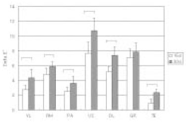
Figure 4 Total color difference (ΔE*). Bar means statistically significant difference between 15days and 30days (p < 0.05)
Reference
-
1. Rosenstiel SF, Land MF, Crispin BJ. Dental luting agents: A review of the current literature. J Prosthet Dent. 1998. 80:280–301.
Article2. Bowen RL, Argenitar H. Amine accelerators for methacrylate resin systems. J Dent Res. 1971. 50:923–928.
Article3. Asmussen E. Factors affecting the color stability of restorative resins. Acta Odontol Scand. 1983. 41:11–18.
Article4. Um CM, Ruyter IE. Staining of resin-based veneering materials with coffee and tea. Quintessence Int. 1991. 22:377–386.5. Hekimoğlu C, Anil N, Etikan I. Effect of Accelerated Aging on the Color Stability of Cemented Laminate Veneers. Int J Prosthodont. 2000. 13:29–33.6. Folwaczny M, Loher C, Mehl A, Kunzelmann KH, Hinkel R. Tooth-Colored Filling Materials for the Restoration of Cervical Lesions: A 24-Month Follow-Up Study. Oper Dent. 2000. 25:251–258.7. Iazzetti G, Burgess JO, Gardiner D, Ripps A. Color stability of Fluoride-Containing Restorative Materials. Oper Dent. 2000. 25:520–525.8. Seghi RR, Gritz MD, Kim J. Colorimetric changes in composites resulting from visible light initiated polymerization. Dent Mater. 1990. 6:133–137.
Article9. Satou N, Khan AM, Matsumae I, Satou J, Shintani H. In vitro color change of composite resins. Dent Mater. 1989. 5:384–389.10. Asmussen E. An accelerated test for color stability of restorative resins. Acta Odontol Scand. 1981. 39:329–332.
Article11. Peutzfeldt A, Asmussen PA. Color stability of three composite resins in the inlay/onlay technique. Scand J Dent Res. 1990. 98:257–260.12. Gross MD, Moser JB. A colorimetric study of coffee and tea staining of four composite resins. J Oral Rehabil. 1977. 4:311–322.
Article13. Wozniak WT, Muller TP, Silverman R, Moser JB. Photographic assessment of color changes in cold and heat-cure resins. J Oral Rehabil. 1981. 8:333–339.
Article14. Noie F, O Keefe KL, Powers JM. Color stability of resin cements after accelerated aging. Int J Prosthodont. 1995. 8:51–55.15. Berrong JM, Weed RM, Schwartz IS. Color stability of selected dual-cured composite resin cement. J Prosthodont. 1993. 2:24–27.16. Setz J, Engel E. In vivo color stability of resin-veneered telescopic dentures: A double blind pilot study. J Prosthet Dent. 1997. 77:486–491.
Article17. Dietschi D, Campanile G, Holz J, Meyer JM. Comparison of the color stability of ten new-generation composites: An in vitro study. Dent Mater. 1994. 10:353–362.
Article18. Abu-Bakr N, Han L, Okamoto A, Iwaku M. Color Stability of Compomer after Immersion in Various Media. J Esthet Dent. 2000. 12:258–263.
Article19. Eliades T, Gioka C, Heim M, Eliades G, Makou M. Color stability of Orthodontic adhesive resins. Angle Orthod. 2004. 74:391–393.20. Koishi Y, Tanoue N, Atsuta M, Matsumura H. Influence of visible-light exposure on colour stability of current dual-curable luting composites. J Oral Rehabil. 2002. 29:387–393.
Article21. Duke ES, Trevino DF. A resin-modified glass ionomer restorative: three-year clinical results. J Indiana Dent Assoc. 1998. 77:13–16.22. Douglas RD. Color stability of new-generation indirect resins for prosthodontic application. J Prosthet Dent. 2000. 83:166–170.
Article23. Rosenstiel SF, Land MF, Crispin BJ. Dental luting agents: A review of the current literature. J Prosthet Dent. 1998. 80:280–301.
Article24. Eldiwany M, Friedl KH, Powers JM. Color stability of light-cured and post cured composites. Am J Dent. 1995. 8:179–181.25. Hwang IN, Oh WM. An Accelerated Test for Color Stability and Opacity Change of Light Curing Composite Resins. J Korean Acad Conserv Dent. 1993. 18:215–226.26. Bergman B, Marklund S, Nilson H, Hendlund SO. An intraindividual clinical comparison of 2 metal-ceramic systems. Int J Prosthodont. 1999. 12:444–447.
- Full Text Links
-
- Actions
-
Cited
- CITED
-
- Close
- Share
-
- Similar articles
-
- Effect of Accelerated Aging on the Color Stability of Dual-Cured Self-Adhesive Resin Cements
- The color stability of aesthetic restorative materials resulting from accelerated aging
- A study on the color stability of resin cements luted for porcelain laminate veneer
- Color stability of current prosthetic composites under accelerated aging and Immersion in a coffee solution
- The study on the color stability of resin cement used in all ceramic crown
