Investig Magn Reson Imaging.
2015 Sep;19(3):162-167. 10.13104/imri.2015.19.3.162.
Quantitative Analysis of the Facial Nerve Using Contrast-Enhanced Three Dimensional FLAIR-VISTA Imaging in Pediatric Bell's Palsy
- Affiliations
-
- 1Department of Radiology, Chungnam National University Hospital, Daejeon, Korea. sunkyou@cnuh.co.kr
- 2Department of Radiology, Kyungpook National University Medical Center, Daegu, Korea.
- 3Department of Radiology, Ewha Womans University Mokdong Hospital, Seoul, Korea.
- KMID: 2175609
- DOI: http://doi.org/10.13104/imri.2015.19.3.162
Abstract
- PURPOSE
To evaluate the usefulness of quantitative analysis of the facial nerve using contrast-enhanced three-dimensional (CE 3D) fluid-attenuated inversion recovery-volume isotopic turbo spin echo acquisition (FLAIR-VISTA) for the diagnosis of Bell's palsy in pediatric patients.
MATERIALS AND METHODS
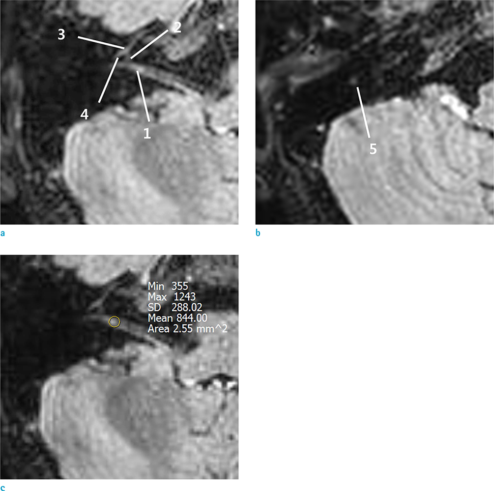
Twelve patients (24 nerves) with unilateral acute facial nerve palsy underwent MRI from March 2014 through March 2015. The unaffected sides were included as a control group. First, for quantitative analysis, the signal intensity (SI) and relative SI (RSI) for canalicular, labyrinthine, geniculate ganglion, tympanic, and mastoid segments of the facial nerve on CE 3D FLAIR images were measured using regions of interest (ROI). Second, CE 3D FLAIR and CE T1-SE images were analyzed to compare their diagnostic performance by visual assessment (VA). The sensitivity, specificity, and accuracy of RSI measurement and VA were compared.
RESULTS
The absolute SI of canalicular and mastoid segments and the sum of the five mean SI (total SI) were higher in the palsy group than in the control group, but with no significant differences. The RSI of the canalicular segment and the total SI were significantly correlated with the symptomatic side (P = 0.028 and 0.015). In 11/12 (91.6%) patients, the RSI of total SI resulted in accurate detection of the affected side. The sensitivity, specificity, and accuracy for detecting Bell's palsy were higher with RSI measurement than with VA of CE 3D FLAIR images, while those with VA of CE T1-SE images were higher than those with VA of CE 3D FLAIR images.
CONCLUSION
Quantitative analysis of the facial nerve using CE 3D FLAIR imaging can be useful for increasing the diagnostic performance in children with Bell's palsy when difficult to diagnose using VA alone. With regard to VA, the diagnostic performance of CE T1-SE imaging is superior to that of CE 3D FLAIR imaging in children. Further studies including larger populations are necessary.
Keyword
MeSH Terms
Figure
Reference
-
1. Baugh RF, Basura GJ, Ishii LE, et al. Clinical practice guideline: Bell's palsy. Otolaryngol Head Neck Surg. 2013; 149:S1–S27.2. Pavlou E, Gkampeta A, Arampatzi M. Facial nerve palsy in childhood. Brain Dev. 2011; 33:644–650.3. Shargorodsky J, Lin HW, Gopen Q. Facial nerve palsy in the pediatric population. Clin Pediatr (Phila). 2010; 49:411–417.4. Gebarski SS, Telian SA, Niparko JK. Enhancement along the normal facial nerve in the facial canal: MR imaging and anatomic correlation. Radiology. 1992; 183:391–394.5. Hong HS, Yi BH, Cha JG, et al. Enhancement pattern of the normal facial nerve at 3.0 T temporal MRI. Br J Radiol. 2010; 83:118–121.6. Hyun D, Lim HK, Park JW, et al. Enhancement pattern of the normal facial nerve on three - dimensional (3D) - fluid attenuated inversion recovery (FLAIR) sequence at 3.0 T MR units. J Korean Soc Magn Reson Med. 2012; 16:25–30.7. Lim HK, Lee JH, Hyun D, et al. MR diagnosis of facial neuritis: diagnostic performance of contrast-enhanced 3D-FLAIR technique compared with contrast-enhanced 3D-T1-fast-field echo with fat suppression. AJNR Am J Neuroradiol. 2012; 33:779–783.8. Kress B, Griesbeck F, Stippich C, Bahren W, Sartor K. Bell palsy: quantitative analysis of MR imaging data as a method of predicting outcome. Radiology. 2004; 230:504–509.9. Song MH, Kim J, Jeon JH, et al. Clinical significance of quantitative analysis of facial nerve enhancement on MRI in Bell's palsy. Acta Otolaryngol. 2008; 128:1259–1265.10. Burmeister HP, Baltzer PA, Volk GF, et al. Evaluation of the early phase of Bell's palsy using 3 T MRI. Eur Arch Otorhinolaryngol. 2011; 268:1493–1500.11. Yun SJ, Ryu CW, Jahng GH, et al. Usefulness of contrast-enhanced 3-dimensional T1-VISTA in the diagnosis of facial neuritis: comparison with contrast-enhanced T1-TSE. J Neuroradiol. 2015; 42:93–98.12. Kim IS, Shin SH, Kim J, Lee WS, Lee HK. Correlation between MRI and operative findings in Bell's palsy and Ramsay Hunt syndrome. Yonsei Med J. 2007; 48:963–968.13. Seok JI, Lee DK, Kim KJ. The usefulness of clinical findings in localising lesions in Bell's palsy: comparison with MRI. J Neurol Neurosurg Psychiatry. 2008; 79:418–420.14. Fukuoka H, Hirai T, Okuda T, et al. Comparison of the added value of contrast-enhanced 3D fluid-attenuated inversion recovery and magnetization-prepared rapid acquisition of gradient echo sequences in relation to conventional postcontrast T1-weighted images for the evaluation of leptomeningeal diseases at 3T. AJNR Am J Neuroradiol. 2010; 31:868–873.
- Full Text Links
- Actions
-
Cited
- CITED
-
- Close
- Share
- Similar articles
-
- How Long Could the Enhancement of Facial Nerve Last in Bell’s Palsy?
- Bilateral Simultaneous Bell's Palsy-Two Case Studies
- Association of the Prognosis and the Facial Nerve Enhancement in Gadolinium Enhanced MRI in Patients with Bell's Palsy
- Clinical Implication of Magnetic Resonance Imaging in Bell's Palsy
- Serious Neurological Disorders That Mimic Bell’s Palsy: A 10-Year Experience


