A Case of Congenital Leukemia Cutis
- Affiliations
-
- 1Department of Dermatology, College of Medicine, Hallym University, Seoul, Korea. dermlee@ yahoo.co.kr
- KMID: 2172071
- DOI: http://doi.org/10.5021/ad.2009.21.1.66
Abstract
- Congenital leukemia is a rare disease that develops from birth to 6 weeks of life. Leukemia cutis involves cutaneous infiltration by leukemic cells and is an unusual manifestation of leukemia, and has been documented in 25~30% of patients with congenital leukemia. The authors report a case of congenital leukemia cutis. A newborn male presented with widespread firm dusky red papules and nodules on almost his entire body surface. Skin biopsy specimens confirmed the presence of leukemic infiltrations, and bone marrow cytology was consistent with acute myeloid leukemia of the FAB M5 type.
MeSH Terms
Figure
-
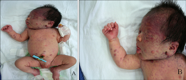
Fig. 1 (A) Multiple scattered dusky red papules and nodules were distributed over the entire body. (B) Diffuse purpuric macules and ecchymoses predominated on scalp, face, and neck.
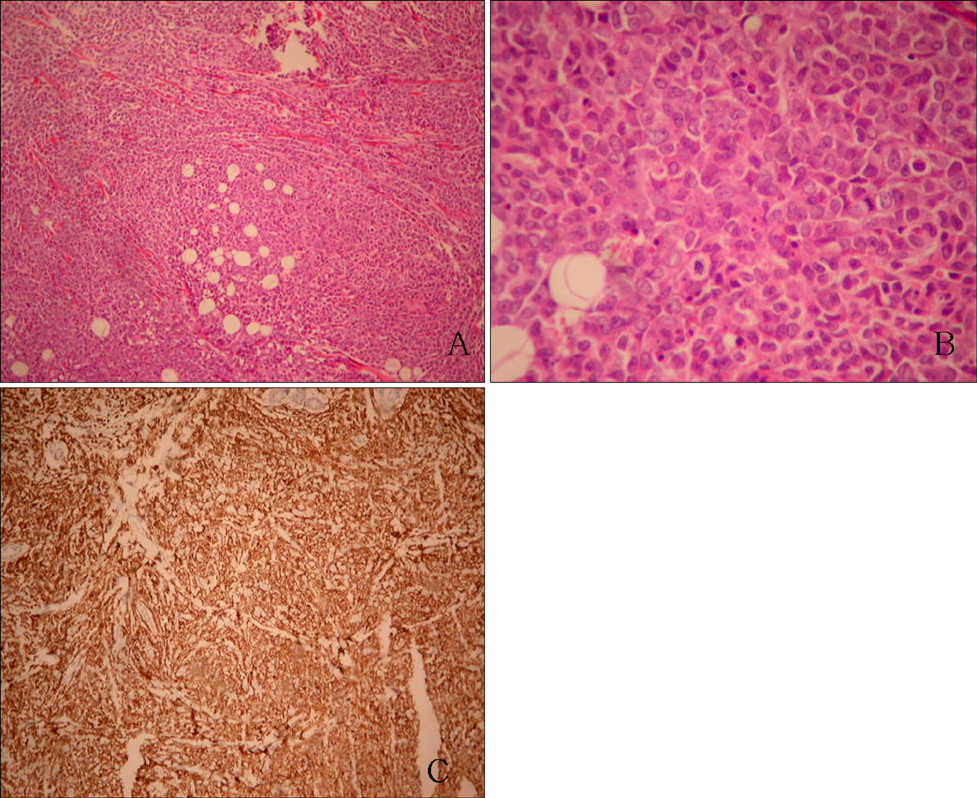
Fig. 2 (A) Biopsy of a skin nodule on the right arm showing dense, diffuse, and atypical infiltration of leukemic cells into dermis and subcutaneous fat (Hematoxylin-eosin, ×100). (B) The majority of infiltrating cells were large pleomorphic cells with a round to oval nucleus with one or more discrete nuclei and abundant pale cytoplasm (Hematoxylin-eosin, ×400). (C) Leukemic cells stained positively for leukocyte common antigen (Immunohistochemical staining, ×100).

Fig. 3 (A) Peripheral blood smear showing markedly elevated monoblast and promonocyte numbers. (B), (C) Bone marrow aspiration showing promonocytes and monocytes containing azurophilic granules. (D) Bone marrow biopsy finding showing that the marrow space was packed with leukemic blasts and immature monocytes. (E), (F) The peripheral blood smear was positive for nonspecific esterase (NSE) and neutrophil activating factor (NaF).
Reference
-
1. Landers MC, Malempati S, Tilford D, Gatter K, White C, Schroeder TL. Spontaneous regression of aleukemia congenital leukemia cutis. Pediatr Dermatol. 2005. 22:26–30.
Article2. Zhang IH, Zane LT, Braun BS, Maize J Jr, Zoger S, Loh ML. Congenital leukemia cutis with subsequent development of leukemia. J Am Acad Dermatol. 2006. 54:S22–S27.
Article3. Isaacs H Jr. Fetal and neonatal leukemia. J Pediatr Hematol Oncol. 2003. 25:348–361.
Article4. Lampkin BC. The newborn infant with leukemia. J Pediatr. 1997. 131:176–177.5. Torrelo A, Madero L, Mediero IG, Bano A, Zambrano A. Aleukemic congenital leukemia cutis. Pediatr Dermatol. 2004. 21:458–461.
Article6. Resnik KS, Brod BB. Leukemia cutis in congenital leukemia. Analysis and review of the world literature with report of an additional case. Arch Dermatol. 1993. 129:1301–1306.
Article7. Bresters D, Reus AC, Veerman AJ, van Wering ER, van der Does-van den Berg A, Kaspers GJ. Congenital leukaemia: the Dutch experience and review of the literature. Br J Haematol. 2002. 117:513–524.
Article8. Crist WM, Pui CH. Nelson WE, Behrman RE, Kliegman RM, Arvin AM, editors. The leukemia. Nelson textbook of pediatrics. 1996. 15th ed. Philadelphia: W.B. Saunders;1452–1457.9. de Lacerda JF, do Carmo JA, Guerra ML, de Almeida LS, Fernandes A, de Lacerda JM. Leukemia cutis in acute lymphoblastic leukemia. J Am Acad Dermatol. 1994. 30:1041–1043.
Article10. Hur J, Kin YS, Yu HJ, Kim JS. A case of congenital leukemia cutis. Ann Dermatol. 2008. 20:74–76.
Article11. Chang SE, Koh KJ, Choi JH, Sung KJ, Moon KC, Koh JK. Congenital leukemia with a leukemic infiltration of skin. Korean J Dermatol. 2000. 38:702–704.12. Ro YS, Moon DG, Lee CW, Han HG, Lee H, Choi JK. A case of congenital leukemia cutis. Korean J Dermatol. 2000. 38:1089–1093.13. Bayoumy M, Wynn T, Jamil A, Kahwash S, Klopfenstein K, Ruymann F. Prenatal presentation supports the in utero development of congenital leukemia: a case report. J Pediatr Hematol Oncol. 2003. 25:148–152.
Article14. Cimino G, Lo Coco F, Biondi A, Elia L, Luciano A, Croce CM, et al. ALL-1 gene at chromosome 11q23 is consistently altered in acute leukemia of early infancy. Blood. 1993. 82:544–546.
Article15. Loh ML, Matthay KK. Taeusch HW, Ballard RA, Gleason CA, editors. Congenital malignant disorders. Avery's diseases of the newborn. 2005. 8th ed. Philadelphia: Elsevier Saunders;1450.
Article16. Longacre TA, Smoller BR. Leukemia cutis. Analysis of 50 biopsy-proven cases with an emphasis on occurrence in myelodysplastic syndromes. Am J Clin Pathol. 1993. 100:276–284.
Article17. Su WP. Clinical, histopathologic, and immunohistochemical correlations in leukemia cutis. Semin Dermatol. 1994. 13:223–230.18. Grundy RG, Martinez A, Kempski H, Malone M, Atherton D. Spontaneous remission of congenital leukemia: a case for conservative treatment. J Pediatr Hematol Oncol. 2000. 22:252–255.
Article19. Lampkin BC, Peipon JJ, Price JK, Bove KE, Srivastava AK, Jones MM. Spontaneous remission of presumed congenital acute nonlymphoblastic leukemia (ANLL) in a karyotypically normal neonate. Am J Pediatr Hematol Oncol. 1985. 7:346–351.
- Full Text Links
-
- Actions
-
Cited
- CITED
-
- Close
- Share
-
- Similar articles
-
- A Case of Congenital Leukemia Cutis
- A Case of Leukemia Cutis Associated with B-cell Chronic Lymphocytic Leukemia
- A Case of Aleukemic Leukemia Cutis Occurring During Spontaneous Remission of Acute Myelomonocytic Leukemia
- A Case of Leukemia Cutis Showing Rosacea-like Cutaneous Lesions
- A Case of Leukemia Cutis Presenting with Extensive Nodular Lesions
