Ann Dermatol.
2009 Feb;21(1):42-45. 10.5021/ad.2009.21.1.42.
Aneurysmal Benign Fibrous Histiocytoma with Atrophic Features
- Affiliations
-
- 1Department of Dermatology, Seoul National University College of Medicine, Boramae Medical Center, Seoul, Korea. sycho@snu.ac.kr
- 2Department of Pathology, Seoul National University College of Medicine, Boramae Medical Center, Seoul, Korea.
- 3Department of Plastic Surgery, Seoul National University College of Medicine, Boramae Medical Center, Seoul, Korea.
- KMID: 2172064
- DOI: http://doi.org/10.5021/ad.2009.21.1.42
Abstract
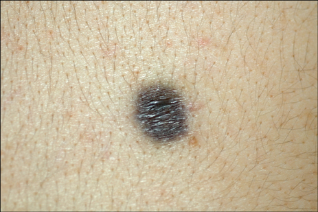
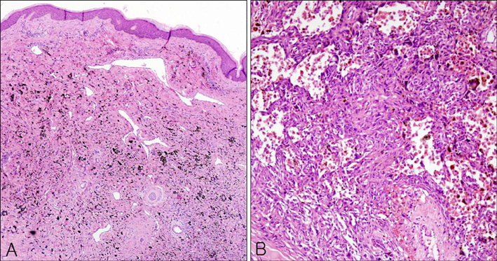
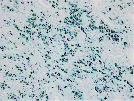
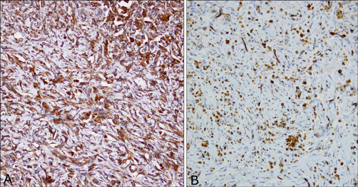
- Aneurysmal benign fibrous histiocytoma is an uncommon pathologic variant of dermatofibroma. In addition to the features of a typical dermatofibroma, it has large cleft-like or cavernous blood-filled spaces with numerous hemosiderin pigments. It should be differentiated from angiomatoid malignant fibrous histiocytoma, malignant melanoma, and vascular tumors such as Kaposi's sarcoma and angiosarcoma. Atrophic dermatofibroma is also a rare variant of dermatofibroma, and the combination of aneurysmal and atrophic features is rarer still. We report a case of aneurysmal benign fibrous histiocytoma with atrophic features in a 27-year-old male who had a grayish-brown atrophic patchy lesion on his back for 2 years.
Keyword
MeSH Terms
Figure
Reference
-
1. Santa Cruz DJ, Kyriakos M. Aneurysmal ("angiomatoid") fibrous histiocytoma of the skin. Cancer. 1981. 47:2053–2061.
Article2. Calonje E, Fletcher CD. Aneurysmal benign fibrous histiocytoma: clinicopathological analysis of 40 cases of a tumour frequently misdiagnosed as a vascular neoplasm. Histopathology. 1995. 26:323–331.
Article3. Goldblum JR, Tuthill RJ. CD34 and factor-XIIIa immunoreactivity in dermatofibrosarcoma protuberans and dermatofibroma. Am J Dermatopathol. 1997. 19:147–153.
Article4. Zelger BW, Zelger BG, Steiner H, Ofner D. Aneurysmal and haemangiopericytoma-like fibrous histiocytoma. J Clin Pathol. 1996. 49:313–318.
Article5. Page EH, Assaad DM. Atrophic dermatofibroma and dermatofibrosarcoma protuberans. J Am Acad Dermatol. 1987. 17:947–950.
Article6. Curco N, Pagerols X, Garcia M, Tarroch X, Vives P. Atrophic dermatofibroma accompanied by aneurysmatic characteristics. J Eur Acad Dermatol Venereol. 2006. 20:331–333.
Article7. Zelger BW, Ofner D, Zelger BG. Atrophic variants of dermatofibroma and dermatofibrosarcoma protuberans. Histopathology. 1995. 26:519–527.
Article8. Kiyohara T, Kumakiri M, Kobayashi H, Ohkawara A, Lao LM. Atrophic dermatofibroma. Elastophagocytosis by the tumor cells. J Cutan Pathol. 2000. 27:312–315.
Article9. Beer M, Eckert F, Schmoeckel C. The atrophic dermatofibroma. J Am Acad Dermatol. 1991. 25:1081–1082.
Article10. Kim EH, Kang HY, Lee ES, Kim YC. Two cases of dermatofibroma with atrophic features. Korean J Dermatol. 2007. 45:305–308.


