Ann Dermatol.
2011 Feb;23(1):89-91. 10.5021/ad.2011.23.1.89.
A Case of Cutaneous Horn Originating from Keratoacanthoma
- Affiliations
-
- 1Department of Dermatology, Soonchunhyang University College of Medicine, Cheonan, Korea. dermsung@schmc.ac.kr
- 2Department of Pathology, Soonchunhyang University College of Medicine, Cheonan, Korea.
- KMID: 2171961
- DOI: http://doi.org/10.5021/ad.2011.23.1.89
Abstract
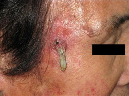
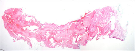
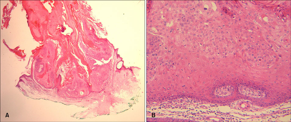
- Cutaneous horn is the clinical description of a hyperproliferation of compact keratin in response to a wide array of underlying benign and malignant pathologic changes. We report here on a case of cutaneous horn that originated from keratoacanthoma in a 76-year-old woman. Grossly, a 2.5x0.7 cm sized yellow-white colored scaly fungating mass from an erythematous nodule was observed on the right temporal area. Histopathologically, it was reported as keratoacanthoma with cutaneous horn. The lesion was totally excised after the diagnosis.
Keyword
Figure
Reference
-
1. Bart RS, Andrade R, Kopf AW. Cutaneous horns. A clinical and histopathologic study. Acta Derm Venereol. 1968. 48:507–515.2. Yu RC, Pryce DW, Macfarlane AW, Stewart TW. A histopathological study of 643 cutaneous horns. Br J Dermatol. 1991. 124:449–452.
Article3. Gould GM, Pyle WL. Anomalies and curiosities in medicine. 1897. 1st ed. Philadelphia: W.B. Saunders;225.4. Bart RS. Andrade R, Gumport SL, Popkin GL, Rees TD, editors. Cutaneous horns. Cancer of the skin: biology-diagnosis-management. 1976. 1st ed. Philadelphia: Saunders;557–572.5. Schosser RH, Hodge SJ, Gaba CR, Owen LG. Cutaneous horns: a histopathologic study. South Med J. 1979. 72:1129–1131.6. Hwang JY, Jeon HD, Lee SY, Lee JS, Chung H, Whang KU. Cutaneous horn arising from keratoacanthoma. Korean J Dermatol. 1998. 36:959–961.7. Kim YJ, Oh ST, Kang H, Park CJ, Park YM, Cho SH, et al. Clinicopathologic study of cutaneous horns. Korean J Dermatol. 2005. 43:359–365.8. Taylor JA. Penile horn. Trans Am Assoc Genitourin Surg. 1945. 37:101–108.9. Koh HK, Bhawan J. Moschella SL, Hurley HJ, editors. Tumor of the skin. Dermatology. 1992. 3rd ed. Philadelphia: WB Saunders;1728–1729.10. Kingman J, Callen JP. Keratoacanthoma. A clinical study. Arch Dermatol. 1984. 120:736–740.
Article11. Rapaport J. Giant keratoacanthoma of the nose. Arch Dermatol. 1975. 111:73–75.
Article12. Ko NY, Park JH, Son SW, Kim IH. Treatment of keratoacanthoma with 5% imiquimod cream. Ann Dermatol. 2006. 18:14–17.
Article13. Schwartz RA. Keratoacanthoma. J Am Acad Dermatol. 1994. 30:1–19.
Article14. Griffiths RW. Keratoacanthoma observed. Br J Plast Surg. 2004. 57:485–501.
Article15. Jasnoch V, Ernst K, Hundeiker M. Rare keratoacanthoma variants. Hautarzt. 1995. 46:244–249.


