Ann Dermatol.
2015 Oct;27(5):608-611. 10.5021/ad.2015.27.5.608.
Preceding Annular Skin Lesions in a Patient with Hemophagocytic Lymphohistiocytosis
- Affiliations
-
- 1Department of Dermatology, Seoul St. Mary's Hospital, College of Medicine, The Catholic University of Korea, Seoul, Korea. yymmpark6301@hotmail.com
- KMID: 2171463
- DOI: http://doi.org/10.5021/ad.2015.27.5.608
Abstract
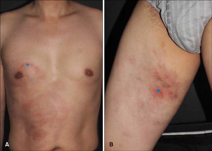
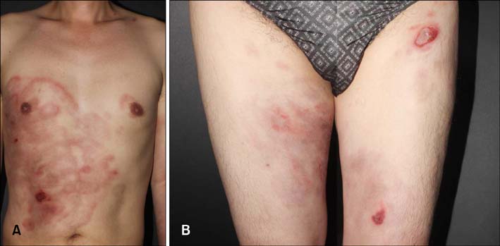
- The cutaneous manifestations of hemophagocytic lymphohistiocytosis (HLH) are variable and nonspecific. A 42-year-old man presented with multiple annular, erythematous patches on the trunk for 3 months. Two months later, he presented with bullae along with high fever. The laboratory examination showed pancytopenia, hypertriglyceridemia, and hypofibrinogenemia. The bone marrow biopsy specimen showed an active hemophagocytosis. On the basis of these findings, a diagnosis of HLH was concluded. After five cycles of chemotherapy, his skin lesion completely resolved. Taking the results together, we suggest that annular skin lesion can be added to the list of cutaneous manifestations of HLH.
MeSH Terms
Figure
Reference
-
1. Morrell DS, Pepping MA, Scott JP, Esterly NB, Drolet BA. Cutaneous manifestations of hemophagocytic lymphohistiocytosis. Arch Dermatol. 2002; 138:1208–1212.
Article2. Gupta S, Weitzman S. Primary and secondary hemophagocytic lymphohistiocytosis: clinical features, pathogenesis and therapy. Expert Rev Clin Immunol. 2010; 6:137–154.
Article3. Fardet L, Galicier L, Vignon-Pennamen MD, Regnier S, Noguera ME, de Labarthe A, et al. Frequency, clinical features and prognosis of cutaneous manifestations in adult patients with reactive haemophagocytic syndrome. Br J Dermatol. 2010; 162:547–553.
Article4. Henter JI, Horne A, Aricó M, Egeler RM, Filipovich AH, Imashuku S, et al. HLH-2004: Diagnostic and therapeutic guidelines for hemophagocytic lymphohistiocytosis. Pediatr Blood Cancer. 2007; 48:124–131.
Article5. DeWitt CA, Buescher LS, Stone SP. Cutaneous manifestations of internal malignant disease: cutaneous paraneoplastic syndromes. In : Goldsmith L, Katz S, Gilchrest B, Paller A, Leffell D, Wolff K, editors. Fitzpatrick's dermatology in general medicine. 8th ed. New York: McGraw-Hill;2012. p. 1880–1892.6. Drago F, Romagnoli M, Loi A, Rebora A. Epstein-Barr virus-related persistent erythema multiforme in chronic fatigue syndrome. Arch Dermatol. 1992; 128:217–222.
Article7. Valeyrie-Allanore L, Raujear JC. Epidermal necrolysis (stevens-Johnson syndrome and toxic epidermal necrolysis). In : Goldsmith L, Katz S, Gilchrest B, Paller A, Leffell D, Wolff K, editors. Fitzpatrick's dermatology in general medicine. 8th ed. New York: McGraw-Hill;2012. p. 439–446.
- Full Text Links
- Actions
-
Cited
- CITED
-
- Close
- Share
- Similar articles
-
- Multiple Ecthyma Gangrenosum in a Hemophagocytic Lymphohistiocytosis Patient
- Hemophagocytic Lymphohistiocytosis Manifesting as a Purpuric Patch
- Chronic Graft-Versus-Host Disease Mimicking Psoriasis in a Patient with Hemophagocytic Lymphohistiocytosis
- Two Cases of Hemophagocytic Lymphohistiocytosis Following Kikuchi's Disease
- A Case of Hemophagocytic Lymphohistiocytosis Presenting with Neck Mass in a Child


