Infect Chemother.
2010 Dec;42(6):431-433. 10.3947/ic.2010.42.6.431.
A Case of Duodenal Ulcer Due to Coinfection with Strongyloides stericoralis and Cytomegalovirus
- Affiliations
-
- 1Department of Internal Medicine, Gyeongsang National University School of Medicine, Jinju, Korea. ttezebae@gmail.com
- 2Department of Parasitology, Gyeongsang National University School of Medicine, Jinju, Korea.
- 3Department of Pathology, Gyeongsang National University School of Medicine, Jinju, Korea.
- 4Gyeongsang Institute of Health Science, Gyeongsang National University School of Medicine, Jinju, Korea.
- KMID: 2170327
- DOI: http://doi.org/10.3947/ic.2010.42.6.431
Abstract
- Gastrointestinal strongyloidiasis and Cytomegalovirus infection mostly occur in patients receiving cancer chemotherapy, undergoing immunosuppressive therapy after organ transplantation, and suffering from acquired immune deficiency syndrome. A 56-year-old man was admitted to the hospital because of abdominal pain and constipation. He had a 10-year history of chronic obstructive pulmonary disease and has been treated intermittently with systemic steroid. The gastroduodenoscopic examination showed a single ulcer on the duodenal bulb and microscopic finding of the biopsy specimens from the ulcer revealed Strongyloides stercoralis and cytomegalovirus immunohistochemical stain positive intranuclear inclusion body on the mucosal surface. The patient was successfully treated with albendazole and ganciclovir.
MeSH Terms
-
Abdominal Pain
Acquired Immunodeficiency Syndrome
Albendazole
Biopsy
Coinfection
Constipation
Cytomegalovirus
Cytomegalovirus Infections
Duodenal Ulcer
Ganciclovir
Humans
Intranuclear Inclusion Bodies
Middle Aged
Organ Transplantation
Pulmonary Disease, Chronic Obstructive
Steroids
Stress, Psychological
Strongyloides
Strongyloides stercoralis
Strongyloidiasis
Transplants
Ulcer
Albendazole
Ganciclovir
Steroids
Figure
-
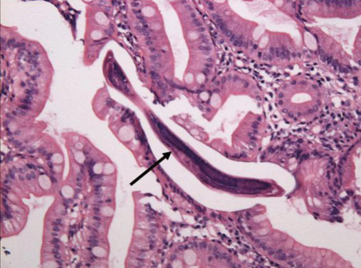
Figure 1 Microscopic appearance of Strongyloides stercoralis from duodenal biopsy specimen. A filariform larva (arrow) is seen in the lumen of an intestinal gland (H&E stain, ×200).
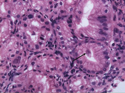
Figure 2 Microscopic appearance of an intranuclear inclusion body from duodenal biopsy specimen. Several glandular epithelial cells (arrow) are enlarged and protrude into the glandular lumen. The nuclei of the cells contain large amphophilic viral inclusions (H&E stain, ×200).
Reference
-
1. Choi SI, Hong SW, Lee KG. Hyperinfection syndrome with Strongyloides stercoralis: report of a case. Korean J Pathol. 1989. 23:359–364.2. Kim J, Joo HS, Kim DH, Lim H, Kang YH, Kim MS. A case of gastric stronglyoidiasis in a Korean patient. Korean J Parasitol. 2003. 41:63–67.
Article3. Hwang IR, Kim JH, Lee KJ, Shin YJ, Yoo BM, Kim YS, Hahm KB, Cho SW. Two cases of CMV esophagitis and proctitis. Korean J Gastrointest Endosc. 1998. 18:763–768.4. Song IS, Choi KW, Kim CY, Jung HC, Kim TH, Kim YS, Kim JS, Lee CH. Efficacy of the treatment with antiviral agent in the cytomegalovirus infection of the gastrointestinal tract. Korean J Gastroenterol. 1998. 32:184–195.5. Wang BY, Krishnan S, Isenberg HD. Mortality associated with concurrent strongyloidosis and cytomegalovirus infection in a patient on steroid therapy. Mt Sinai J Med. 1999. 66:128–132.6. Gupta S, Jain A, Fanning TV, Couriel DR, Jimenez CA, Eapen GA. An unusual cause of alveolar hemorrhage post hematopoietic stem cell transplantation: a case report. BMC Cancer. 2006. 6:87.
Article7. Keiser PB, Nutman TB. Strongyloides stercoralis in the immunocompromised population. Clin Microbiol Rev. 2004. 17:208–217.
Article8. Yoon DH, Yang SJ, Kim JS, Hong ST, Chai JY, Lee SH, Chi JG. A case of fatal malabsorption syndrome caused by strongyloidiasis complicated with isosporiasis and human cytomegalovirus infection. Korean J Parasitol. 1992. 30:53–58.
Article9. Tsai HC, Lee SS, Liu YC, Lin WR, Huang CK, Chen YS, Wann SR, Tsai TH, Lin HH, Yen MY, Yen CM, Chen ER. Clinical manifestations of strongyloidiasis in southern Taiwan. J Microbiol Immunol Infect. 2002. 35:29–36.10. Rhee JC, Choi KW, Lee HY, Koh KC, Paik SW, Lee JK, Lee KT, Lee JH, Lee SH, Kang IK, Hyun JG, Rhee PL, Kim JJ, Oh YR, Chae JI. A case of gastric strongyloidiasis diagnosed by endoscopic biopsy. Korean J Gastrointest Endosc. 1999. 19:249–253.
- Full Text Links
-
- Actions
-
Cited
- CITED
-
- Close
- Share
-
- Similar articles
-
- Upper Gastrointestinal Hemorrhage from Esophageal Ulcer Caused by Coinfection of Herpes Simplex Virus and Cytomegalovirus in Immunocompromised Patient
- A case of strongyloidiasis accompanied by duodenal ulcer
- A Case of Cutaneous Cytomegalovirus Infection in a Renal Transplant Patient
- A Case of Resolution of Duodenal Obstruction Caused by Duodenal Ulcer after the Eradication of Helicobacter pylori Infection
- Three Cases of Coexistence of Gastric Cancer and Duodenal Ulcer
