Endocrinol Metab.
2010 Sep;25(3):236-239. 10.3803/EnM.2010.25.3.236.
A Case of Pheochromocytoma Presented with Cardiogenic Shock and Followed by Spontaneous Remission
- Affiliations
-
- 1Department of Internal Medicine, Sahmyook-Seoul Hospital, Seoul, Korea. 1022p@hanmail.net
- 2Department of Radiology, Sahmyook-Seoul Hospital, Seoul, Korea.
- KMID: 2169069
- DOI: http://doi.org/10.3803/EnM.2010.25.3.236
Abstract
- Pheochromocytoma is derived from the chromaffin cells and patients with pheochromocytoma present with several signs and symptoms by producing, storing and secreting catecholamine. Spontaneous rupture or necrosis of pheochromocytoma is extremely rare, but it can be lethal because of the dramatic change in the circulation such as an acute abdominal emergency or shock. Spontaneous remission of the clinical symptoms due to necrosis of the pheochromocytoma is rare. We describe such a case that presented with cardiogenic shock due to extensive necrosis of the pheochromocytoma and this was followed by spontaneous remission of the clinical symptoms without removal of the pheochromocytoma.
MeSH Terms
Figure
-

Fig. 1 A. Abdominal computed tomography shows about 4 × 4 cm sized, relatively well-defined low density mass lesion with peripheral wall enhancement (arrow) in the left adrenal gland. B. After three months, the size of relatively well-defined low density mass lesion with peripheral wall subtle enhancement (about 1.5 × 1.5 cm) is decreased (arrow).
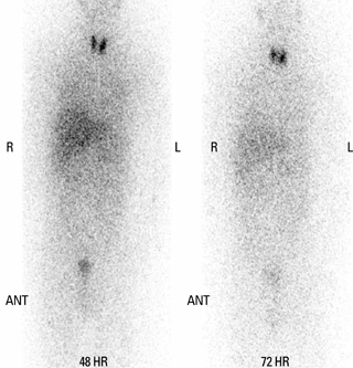
Fig. 2 131I-MIBG scan shows no demonstrable abnormal finding.
Reference
-
1. Larsen PR, Kronenberg HM, Melmed S, Polonsky KS. Williams textbook of endocrinology. 2008. 11th ed. Philadelphia: Saunders;507–522.2. Shin DH, Kim SG, Kim DR, Kim NH, Choi KM, Baik SY, Choi DS, Suh SO. Clinical study of the pheochromocytoma. J Korean Soc Endocrinol. 2002. 17:554–563.3. Ong KL, Tan TH. Ruptured phaeochromocytoma--a rare differential diagnosis of acute abdomen. Singapore Med J. 1996. 37:113–114.4. Kobayashi T, Iwai A, Takahashi R, Ide Y, Nishizawa K, Mitsumori K. Spontaneous rupture of adrenal pheochromocytoma: review and analysis of prognostic factors. J Surg Oncol. 2005. 90:31–35.5. Schifferdecker B, Kodali D, Hausner E, Aragam J. Adrenergic shock--an overlooked clinical entinity? Cardiol Rev. 2005. 13:69–72.6. Terachi T, Terai A, Yoshida S, Yokota K, Fukunaga M. Spontaneous rupture of adrenal pheochromocytoma: a case report. Urol Int. 1989. 44:235–237.7. Van way CW 3rd, Faraci RP, Cleveland HC, Foster JF, Scott HW Jr. Hemorrhagic necrosis of pheochromocytoma associated with phentolamine administration. Ann Surg. 1976. 184:26–30.8. Ejerblad S, Hemmingsson A. Haemorrhage into a pheochromocytoma in an anticoagulant-treated patient. Acta Chir Scand. 1981. 147:497–500.9. Raab W, de Paula e Silva P, Starcheska YK. Adrenergic and cholinergic influences on the dynamic cycle of the normal human heart. Cardiologia. 1958. 33:350–364.10. Szakacs JE, Mehlman B. Pathologic changes induced by 1-norepinephrine: quantitative aspects. Am J Cardiol. 1960. 5:619–627.11. Jiang JP, Downing SE. Catecholamine cardiomyopathy: review and analysis of pathogenetic mechanisms. Yale J Biol Med. 1990. 63:581–591.12. Sardesai SH, Mourant AJ, Sivathandon Y, Farrow R, Gibbons DO. Phaeochromocytoma and catecholamine induced cardiomyopathy presenting as heart failure. Br Heart J. 1990. 63:234–237.13. Yun KH, Lee KH, Rhee BH, Chae JK, Kim WH, Ko JK. A case of pheochromocytoma presented with life: threatening cardiogenic shock. Korean Circ J. 2001. 31:1075–1080.14. Zanin L, Rossi G, Poletti A, Piotto A, Chiesura-Corona M, Pessina AC. Necrosis of a phaeochromocytoma associated with spontaneous remission of diabetes and hypertension. Clin Endocrinol (Oxf). 1993. 39:613–617.15. Terai A, Terachi T, Yoshida S, Kadota K. Pheochromocytoma presenting as shock and followed by spontaneous remission. Urol Int. 1989. 44:58–60.
- Full Text Links
-
- Actions
-
Cited
- CITED
-
- Close
- Share
-
- Similar articles
-
- A Case of Pheochromocytoma Presented with Severe Left Ventricular Dysfunction Combined with Cardiogenic Shock after Caesearian Section Delivery Responding to Treatment of Extracorporeal Membrane Oxygenation
- Catastrophic catecholamine-induced cardiomyopathy rescued by extracorporeal membrane oxygenation in recurrent malignant pheochromocytoma
- A Case of Pheochromocytoma Presented with Cardiogenic Shock
- Pheochromocytoma-induced cardiogenic shock successfully treated by extracorporeal circulation
- A Case of Pheochromocytoma Presented with Life: Threatening Cardiogenic Shock
