Involvement of Protein Kinase C-delta in Vascular Permeability in Acute Lung Injury
- Affiliations
-
- 1Department of Internal Medicine, Ulsan University Hospital, School of Medicine, University of Ulsan, Ulsan 44033, Korea.
- 2Department of Thoracic Surgery, Ulsan University Hospital, University of Ulsan College of Medicine, Ulsan 44033, Korea.
- 3Department of Anesthesiology and Pain Medicine, Ulsan University Hospital, University of Ulsan College of Medicine, Ulsan 44033, Korea.
- 4School of Biological Sciences, University of Ulsan, Ulsan 44610, Korea. bkwon@ulsan.ac.kr
- 5Biomedical Research Center, Ulsan University Hospital, University of Ulsan College of Medicine, Ulsan 44033, Korea. hrcho@uuh.ulsan.kr
- 6Department of Surgery, Ulsan University Hospital, University of Ulsan College of Medicine, Ulsan 44033, Korea.
- KMID: 2168048
- DOI: http://doi.org/10.4110/in.2015.15.4.206
Abstract
- Pulmonary edema is a major cause of mortality due to acute lung injury (ALI). The involvement of protein kinase C-delta (PKC-delta) in ALI has been a controversial topic. Here we investigated PKC-delta function in ALI using PKC-delta knockout (KO) mice and PKC inhibitors. Our results indicated that although the ability to produce proinflammatory mediators in response to LPS injury in PKC-delta KO mice was similar to that of control mice, they showed enhanced recruitment of neutrophils to the lung and more severe pulmonary edema. PKC-delta inhibition promoted barrier dysfunction in an endothelial cell layer in vitro, and administration of a PKC-delta-specific inhibitor significantly increased steady state vascular permeability. A neutrophil transmigration assay indicated that the PKC-delta inhibition increased neutrophil transmigration through an endothelial monolayer. This suggests that PKC-delta inhibition induces structural changes in endothelial cells, allowing extravasation of proteins and neutrophils.
MeSH Terms
Figure
-
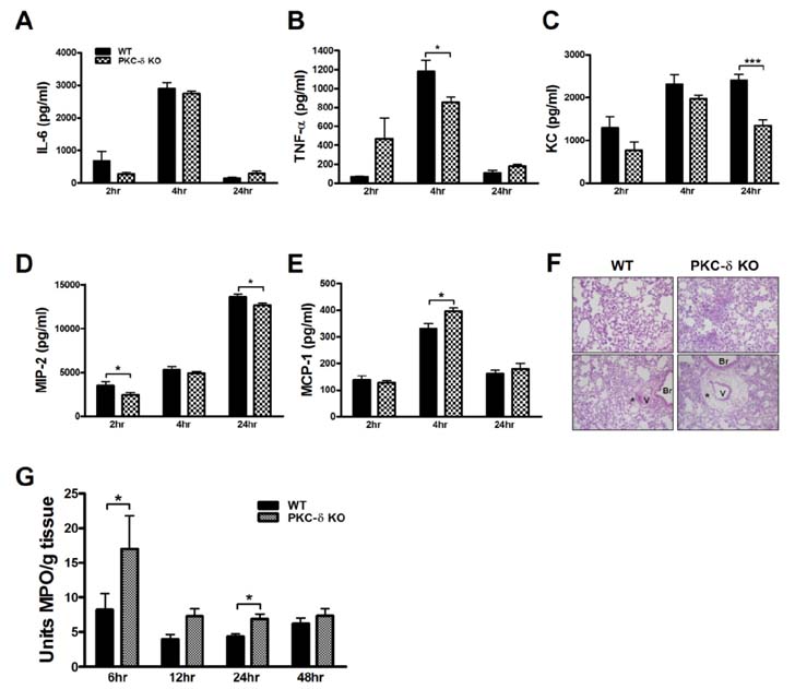
Figure 1 PKC-δ KO mice had no impairment in the production of proinflammatory mediators, but had severe neutrophil infiltration and perivascular edema. (A~E) BAL fluid was harvested at various time points after LPS infusion and levels of proinflammatory cytokines, TNF-α (A) and IL-6 (B), and chemokines, KC (C), MIP-2 (D) and MCP-1 (E), were measured by ELISA. n=4-10 for each group. *p<0.05 and ***p<0.001 between the indicated groups. (F) Lungs were harvested 12 h after LPS infusion and histological analysis was performed. Neutrophil infiltration and perivascular edema were more prominent in PKC-δ KO mice (right column). *perivascular cuff, V: blood vessel, Br: bronchus. Original magnification: ×40 (upper column) and ×200 (lower column). (G) Determination of myeloperoxidase activity in lung tissue. n=7-12 for each group. *p<0.05 and ***p<0.001 between the indicated groups.
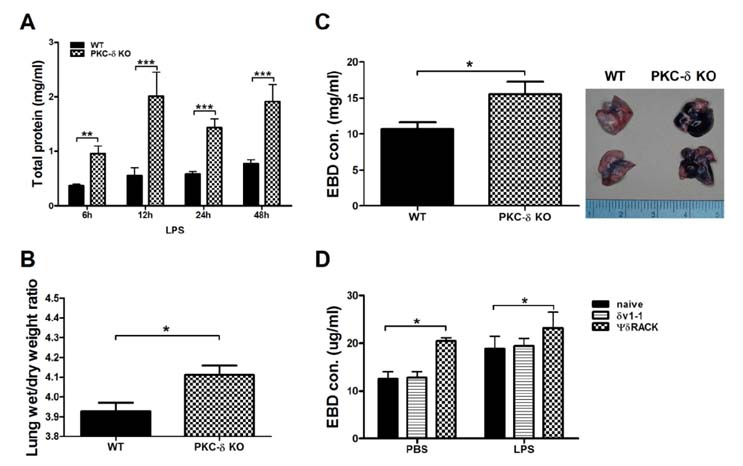
Figure 2 Inhibition of PKC-δ promotes pulmonary edema and vascular permeability. (A) Total protein levels in BAL fluid at various time points after LPS infusion. n=3-14 for each group. *p<0.05, **p<0.01, and ***p<0.001 between the two groups. (B) The lung wet/dry weight ratio at 24 h after LPS infusion. n=12-13 for each group. *p<0.05. (C and D) EBD was injected 4 h after LPS infusion and lungs were harvested 30 min later. Gross observation (left column) and concentrations of EBD in extracted lung tissue (right column). n=12-13 for each group. *p<0.05. (D)δV1-1 or ΨδRACK peptide was intratracheally injected 30 min before LPS infusion. Lungs were harvested 30 min after EBD injection. n=6-8 for each group. *p<0.05 between the indicated groups.
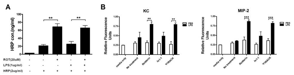
Figure 3 Inhibition of PKC-δ promotes extravasation of proteins and neutrophils through an endothelial monolayer. (A) A confluent monolayer of HUVEC cells was treated with rottlerin 30 min before the addition of LPS. Four hour later, HRP was added to HUVEC cells and extravasation was allowed to proceed for 30 min. n=3 for each group. *p<0.05 and ***p<0.001 between the indicated groups. (B) PKC-δ inhibitors and LPS were treated as described in A and neutrophil transmigration was allowed for 15 h. n=3 for each group. **p<0.01 and ***p<0.001 between the indicated groups.
Reference
-
1. Han S, Mallampalli RK. The acute respiratory distress syndrome: from mechanism to translation. J Immunol. 2015; 194:855–860.
Article2. Siflinger-Birnboim A, Johnson A. Protein kinase C modulates pulmonary endothelial permeability: a paradigm for acute lung injury. Am J Physiol Lung Cell Mol Physiol. 2003; 284:L435–L451.
Article3. Klinger JR, Murray JD, Casserly B, Alvarez DF, King JA, An SS, Choudhary G, Owusu-Sarfo AN, Warburton R, Harrington EO. Rottlerin causes pulmonary edema in vivo: a possible role for PKCδ. J Appl Physiol. 2007; 103:2084–2094.
Article4. Gaudreault N, Perrin RM, Guo M, Clanton CP, Wu MH, Yuan SY. Counter regulatory effects of PKCβII and PKCδ on coronary endothelial permeability. Arterioscler Thromb Vasc Biol. 2008; 28:1527–1533.
Article5. Chichger H, Grinnell KL, Casserly B, Chung CS, Braza J, Lomas-Neira J, Ayala A, Rounds S, Klinger JR, Harrington EO. Genetic disruption of protein kinase Cδ reduces endotoxin-induced lung injury. Am J Physiol Lung Cell Mol Physiol. 2012; 303:L880–L888.
Article6. Miyamoto A, Nakayama K, Imaki H, Hirose S, Jiang Y, Abe M, Tsukiyama T, Nagahama H, Ohno S, Hatakeyama S, Nakayama KI. Increased proliferation of B cells and auto-immunity in mice lacking protein kinase Cdelta. Nature. 2002; 416:865–869.
Article7. Lomas-Neira J, Chung CS, Grutkoski PS, Dunican A, Simms HH, Cioffi WG, Ayala A. Divergent roles of murine neutrophil chemokines in hemorrhage induced priming for acute lung injury. Cytokine. 2005; 31:169–179.
Article8. Neff TA, Guo RF, Neff SB, Sarma JV, Speyer CL, Gao H, Bernacki KD, Huber-Lang M, McGuire S, Hoesel LM, Riedemann NC, Beck-Schimmer B, Zetoune FS, Ward PA. Relationship of acute lung inflammatory injury to Fas/FasL system. Am J Pathol. 2005; 166:685–694.
Article9. Patterson CE, Rhoades RA, Garcia JG. Evans blue dye as a marker of albumin clearance in cultured endothelial monolayer and isolated lung. J Appl Physiol (1985). 1992; 72:865–873.
Article10. Chen L, Hahn H, Wu G, Chen CH, Liron T, Schechtman D, Cavallaro G, Banci L, Guo Y, Bolli R, Dorn GW 2nd, Mochly-Rosen D. Opposing cardioprotective actions and parallel hypertrophic effects of delta PKC and epsilon PKC. Proc Natl Acad Sci U S A. 2001; 98:11114–11119.
Article11. Baudin B, Bruneel A, Bosselut N, Vaubourdolle M. A protocol for isolation and culture of human umbilical vein endothelial cells. Nat Protoc. 2007; 2:481–485.
Article
- Full Text Links
-
- Actions
-
Cited
- CITED
-
- Close
- Share
-
- Similar articles
-
- Effects of Protein Kinase C on Hypoxic Pulmonary Vasoconstriction in Isolated Rat Lungs
- Angiopoietin-1 variant, COMP-Ang1 attenuates hydrogen peroxide-induced acute lung injury
- Role of Protein Kinase C-delta in Atherosclerosis
- Effects of sevoflurane on tight junction protein expression and PKC-alpha translocation after pulmonary ischemia-reperfusion injury
- Morphine and remifentanil-induced cardioprotection: its experimental and clinical outcomes
