Comparison of high-resolution and standard zoom imaging modes in cone beam computed tomography for detection of longitudinal root fracture: An in vitro study
- Affiliations
-
- 1Oro-Maxillofacial Developmental Disease Research Center, Department of Endodontics, Faculty of Dentistry, Guilan University of Medical Sciences, Rasht, Iran.
- 2Department of Maxillofacial Radiology, Oro-Maxillofacial Developmental Disease Research Center, Faculty of Dentistry, Guilan University of Medical Sciences, Rasht, Iran. zahradalili@yahoo.com
- 3Department of Maxillofacial Radiology, Faculty of Dentistry, Guilan University of Medical Sciences, Rasht, Iran.
- 4Vice Chancellor of Research and Technology, Guilan University of Medical Sciences, Rasht, Iran.
- KMID: 2167454
- DOI: http://doi.org/10.5624/isd.2013.43.3.171
Abstract
- PURPOSE
The purpose of this study was to compare the efficacy of two imaging modes in a cone beam computed tomography (CBCT) system in detecting root fracture in endodontically-treated teeth with fiber posts or screw posts by selecting two fields of view.
MATERIALS AND METHODS
In this study, 78 endodontically-treated single canal premolars were included. A post space was created in all of them. Then the teeth were randomly set in one of 6 artificial dental arches. In 39 of the 78 teeth set in the 6 dental arches, a root fracture was intentionally created. Next, a fiber post and a screw post were cemented into 26 teeth having equal the root fractures. High resolution (HiRes) and standard zoom images were provided by a CBCT device. Upon considering the reconstructed images, two observers in agreement with each other confirmed the presence or absence of root fracture. A McNemar test was used for comparing the results of the two modes.
RESULTS
The frequency of making a correct diagnosis using the HiRes zoom imaging mode was 71.8% and in standard zoom was 59%. The overall sensitivity and specificity in diagnosing root fracture in the HiRes mode were 71.79% and 46.15% and in the standard zoom modes were 58.97% and 33.33%, respectively.
CONCLUSION
There were no significant differences between the diagnostic values of the two imaging modes used in the diagnosis of root fracture or in the presence of root canal restorations. In both modes, the most true-positive results were reported in the post space group.
MeSH Terms
Figure
-
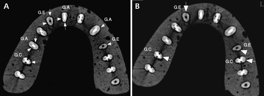
Fig. 1 The axial images of high resolution (A) and standard (B) zoom modes of CBCT images show several root fractures (arrows). The teeth with a fiber post and root fracture (Group A; G.A), with a screw post and root fracture (Group C; G.C), and with a post space and root fracture (Group E; G.E) are seen in each arch set.
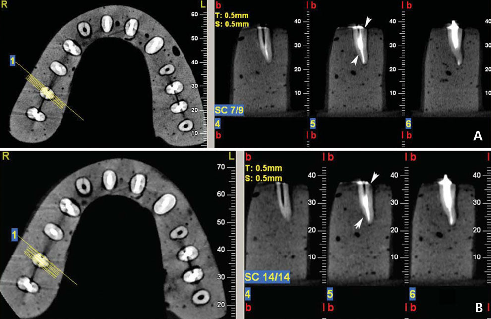
Fig. 2 The axial and cross-sectional images of high resolution (A) and standard (B) zoom modes show the direction of root fracture in a tooth with a screw post and root fracture (arrows).
Cited by 2 articles
-
Assessment of vertical root fracture using cone-beam computed tomography
Ehsan Moudi, Sina Haghanifar, Zahrasadat Madani, Abdolhamid Alhavaz, Ali Bijani, Mohammad Bagheri
Imaging Sci Dent. 2014;44(1):37-41. doi: 10.5624/isd.2014.44.1.37.Effect of titanium and stainless steel posts in detection of vertical root fractures using NewTom VG cone beam computed tomography system
Mahdis Mohammadpour, Neema Bakhshalian, Shahriar Shahab, Shaya Sadeghi, Mona Ataee, Soodeh Sarikhani
Imaging Sci Dent. 2014;44(2):89-94. doi: 10.5624/isd.2014.44.2.89.
Reference
-
1. Rivera EM, Walton RE. Longitudinal toot fractures. In : Torabinejad M, Walton RE, editors. Endodontics; principles and practice. 4th ed. St. Louis: Saunders Elsevier;2009. p. 108–128.2. Meister F Jr, Lommel TJ, Gerstein H. Diagnosis and possible causes of vertical root fracture. Oral Surg Oral Med Oral Pathol. 1980; 49:243–253.3. Peciuliene V, Rimkuviene J. Vertical root fractures in endodontically treated teeth: a clinical survey. Stomatologija. 2004; 6:77–80.4. Hülsmann M, Schäfer E. Problems in endodontics: etiology, diagnosis, and treatment. London: Quintessence;2009. p. 353–371.5. Mora MA, Mol A, Tyndall DA, Rivera EM. In vitro assessment of local computed tomography for the detection of longitudinal tooth fractures. Oral Surg Oral Med Oral Pathol Oral Radiol Endod. 2007; 103:825–829.
Article6. Morfis AS. Vertical root fractures. Oral Surg Oral Med Oral Pathol. 1990; 69:631–635.
Article7. Rud J, Omnell KA. Root fractures due to corrosion. Diagnostic aspects. Scand J Dent Res. 1970; 78:397–403.
Article8. Bargholz C. Vertical tooth and root fractures. In : Hülsmann M, Schäfer E, editors. Problems in endodontics; etiology, diagnosis and treatment. London: Quintessence;2009. p. 353–359.9. Bernardes RA, de Moraes IG, Húngaro Duarte MA, Azevedo BC, de Azevedo JR, Bramante CM. Use of cone-beam volumetric tomography in the diagnosis of root fractures. Oral Surg Oral Med Oral Pathol Oral Radiol Endod. 2009; 108:270–277.
Article10. Hassan B, Metska ME, Ozok AR, van der Stelt P, Wesselink PR. Detection of vertical root fractures in endodontically treated teeth by a cone beam computed tomography scan. J Endod. 2009; 35:719–722.
Article11. Wang P, Yan XB, Lui DG, Zhang WL, Zhang Y, Ma XC. Detection of dental root fractures by using cone-beam computed tomography. Dentomaxillofac Radiol. 2011; 40:290–298.
Article12. Kajan ZD, Taromsari M. Value of cone beam CT in detection of dental root fractures. Dentomaxillofac Radiol. 2012; 41:3–10.
Article13. Metska ME, Aartman IH, Wesselink PR, Özok AR. Detection of vertical root fractures in vivo in endodontically treated teeth by cone-beam computed tomography scans. J Endod. 2012; 38:1344–1347.14. Hassan B, Metska ME, Ozok AR, van der Stelt P, Wesselink PR. Comparison of five cone beam computed tomography systems for the detection of vertical root fractures. J Endod. 2010; 36:126–129.
Article15. Liang X, Jacobs R, Hassan B, Li L, Pauwels R, Corpas L, et al. A comparative evaluation of Cone Beam Computed Tomography (CBCT) and Multi-Slice CT (MSCT) Part I. On subjective image quality. Eur J Radiol. 2010; 75:265–269.16. Katsumata A, Hirukawa A, Okumura S, Naitoh M, Fujishita M, Ariji E, et al. Relationship between density variability and imaging volume size in cone-beam computerized tomographic scanning of the maxillofacial region: an in vitro study. Oral Surg Oral Med Oral Pathol Oral Radiol Endod. 2009; 107:420–425.
Article17. Zhang Y, Zhang L, Zhu XR, Lee AK, Chambers M, Dong L. Reducing metal artifacts in cone-beam CT images by preprocessing projection data. Int J Radiat Oncol Biol Phys. 2007; 67:924–932.
Article18. Özer SY. Detection of vertical root fractures by using cone beam computed tomography with variable voxel sizes in an in vitro model. J Endod. 2011; 37:75–79.19. Dalili Z, Taramsari M, Mousavi Mehr SZ, Salamat F. Diagnostic value of two modes of cone-beam computed tomography in evaluation of simulated external root resorption: an in vitro study. Imaging Sci Dent. 2012; 42:19–24.
Article20. Liedke GS, da Silveira HE, Silveira HL, Dutra V, de Figueiredo JA. Influence of voxel size in the diagnostic ability of cone beam tomography to evaluate simulated external root resorption. J Endod. 2009; 35:233–235.
Article21. Youssefzadeh S, Gahleitner A, Dorffner R, Bernhart T, Kainberger FM. Dental vertical root fractures: value of CT in detection. Radiology. 1999; 210:545–549.
Article
- Full Text Links
-
- Actions
-
Cited
- CITED
-
- Close
- Share
-
- Similar articles
-
- Diagnostic value of two modes of cone-beam computed tomography in evaluation of simulated external root resorption: an in vitro study
- Diagnostic accuracy of cone-beam computed tomography scans with high- and low-resolution modes for the detection of root perforations
- Detection of maxillary second molar with two palatal roots using cone beam computed tomography: a case report
- Three-dimensional imaging modalities in endodontics
- Basic principle of cone beam computed tomography
