Diagnostic value of two modes of cone-beam computed tomography in evaluation of simulated external root resorption: an in vitro study
- Affiliations
-
- 1Department of Maxillofacial Radiology, Dental School, Guilan University of Medical Sciences, Rasht, Iran. zahradalili@yahoo.com
- 2Department of Endodontics, Dental School, Guilan University of Medical Sciences, Rasht, Iran.
- 3Department of Epidemiology, Guilan University of Medical Sciences, Rasht, Iran.
- KMID: 1974403
- DOI: http://doi.org/10.5624/isd.2012.42.1.19
Abstract
- PURPOSE
Field of view and voxel resolution of cone beam computed tomography (CBCT) might affect the diagnostic capability. This study was performed to compare between the standard and HiRes zoom modes in the diagnosis of external root resorption (ERR) using CBCT.
MATERIALS AND METHODS
Sixty three small cavities (0.25 mm depth and 0.5 mm diameter) were simulated on the buccal, lingual, and proximal surfaces at three different levels of 16 roots of teeth. After covering the root with nail varnish, the roots were inserted in the sockets and the model was placed in a water-containing lacuna. CBCT scans were taken in both standard and HiRes zoom modes using NewTom VG (QR srl Company, Verona, Italy). Then, an observer assessed the images to determine the presence or absence of the cavities. This process was repeated by increasing the size and depth of cavities to 0.5 mm depth and 1 mm diameter. Data were analyzed by McNemar test. The sensitivity, specificity, positive predictive value, negative predictive value, and likelihood ratio in evaluation of the simulated cavities were calculated.
RESULTS
There was a significant difference between the two imaging modes in diagnosing the shallow cavities (p=0.02).The sensitivity of the standard zoom in detecting the shallow cavities was lower than that of the HiRes zoom. The likelihood ratio of the HiRes zoom was higher in the diagnosis of both cavity types.
CONCLUSION
This study suggested that a smaller voxel size in the HiRes zoom mode of CBCT is preferred for diagnosis of ERR.
Figure
-
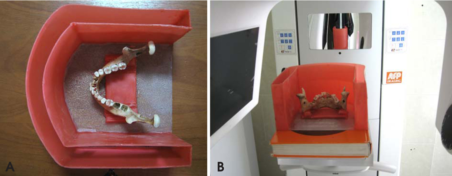
Fig. 1 The photographs present the mandible that mounted in U-shaped water-containing lacuna fixed on a plexy plate and the setting of this complex onto the desk of CBCT device.
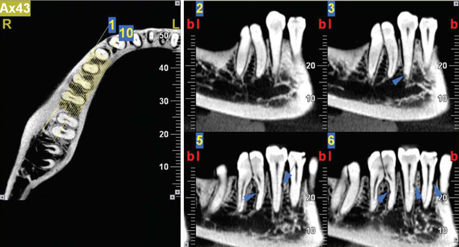
Fig. 2 Mesio-distal cross-sectional images reveal small external root resorption cavities on the right mandibular premolars and first molar tooth in the different locations in the HiRes zoom (arrows).
Cited by 2 articles
-
Comparison of high-resolution and standard zoom imaging modes in cone beam computed tomography for detection of longitudinal root fracture: An in vitro study
Mehran Taramsari, Zahra Dalili Kajan, Parinaz Bashirzadeh, Fatemeh Salamat
Imaging Sci Dent. 2013;43(3):171-177. doi: 10.5624/isd.2013.43.3.171.Diagnostic accuracy of cone-beam computed tomography scans with high- and low-resolution modes for the detection of root perforations
Abbas Shokri, Amir Eskandarloo, Marouf Norouzi, Jalal Poorolajal, Gelareh Majidi, Alireza Aliyaly
Imaging Sci Dent. 2018;48(1):11-19. doi: 10.5624/isd.2018.48.1.11.
Reference
-
1. Hülsmann M, Schäfer E. Problems in endodontics; etiology, diagnosis and treatment. 2009. London: Quintessence;421–434.2. Borg E, Källqvist A, Gröndahl K, Gröndahl HG. Film and digital radiography for detection of simulated root resorption cavities. Oral Surg Oral Med Oral Pathol Oral Radiol Endod. 1998. 86:110–114.
Article3. Chapnick L. External root resorption: an experimental radiographic evaluation. Oral Surg Oral Med Oral Pathol. 1989. 67:578–582.
Article4. Nance RS, Tyndall D, Levin LG, Trope M. Diagnosis of external root resorption using TACT (tuned-aperture computed tomography). Endod Dent Traumatol. 2000. 16:24–28.
Article5. Westphalen VP, Gomes de Moraes I, Westphalen FH, Martins WD, Souza PH. Conventional and digital radiographic methods in the detection of simulated external root resorption: a comparative study. Dentomaxillofac Radiol. 2004. 33:233–235.6. Kravitz LH, Tyndall DA, Bagnell CP, Dove SB. Assessment of external root resorption using digital subtraction radiography. J Endod. 1992. 18:275–284.
Article7. Hintze H, Wenzel A, Andreasen FM, Sewerin I. Digital subtraction radiography for assessment of simulated root resorption cavities. Performance of conventional and reverse contrast modes. Endod Dent Traumatol. 1992. 8:149–154.
Article8. da Silveira HL, Silveira HE, Liedke GS, Lermen CA, Dos Santos RB, de Figueiredo JA. Diagnostic ability of computed tomography to evaluate external root resorption in vitro. Dentomaxillofac Radiol. 2007. 36:393–396.9. Kim E, Kim KD, Roh BD, Cho YS, Lee SJ. Computed tomography as a diagnostic aid for extracanal invasive resorption. J Endod. 2003. 29:463–465.
Article10. Liedke GS, da Silveira HE, Silveira HL, Dutra V, de Figueiredo JA. Influence of voxel size in the diagnostic ability of cone beam tomography to evaluate simulated external root resorption. J Endod. 2009. 35:233–235.
Article11. Patel S, Dawood A, Wilson R, Horner K, Mannocci F. The detection and management of root resorption lesions using intraoral radiography and cone beam computed tomography - an in vivo investigation. Int Endod J. 2009. 42:831–838.12. Hahn W, Fricke-Zech S, Fricke J, Gruber RM, Dullin C, Zapf A, et al. Detection and size differentiation of simulated tooth root defects using flat-panel volume computerized tomography (fpVCT). Oral Surg Oral Med Oral Pathol Oral Radiol Endod. 2009. 107:272–278.
Article13. Nakata K, Naitoh M, Izumi M, Ariji E, Nakamura H. Evaluation of correspondence of dental Computed Tomography imaging to anatomic observation of external root resorption. J Endod. 2009. 35:1594–1597.
Article14. Estrela C, Bueno MR, De Alencar AH, Mattar R, Valladares Neto J, Azevedo BC, et al. Method to evaluate inflammatory root resorption by using cone beam computed tomography. J Endod. 2009. 35:1491–1497.
Article15. Andreasen FM, Sewerin I, Mandel U, Andreasen JO. Radiographic assessment of simulated root resorption cavities. Endod Dent Traumatol. 1987. 3:21–27.
Article16. Goldberg F, De Silvio A, Dreyer C. Radiographic assessment of simulated external root resorption cavities in maxillary incisors. Endod Dent Traumatol. 1998. 14:133–136.
Article
- Full Text Links
-
- Actions
-
Cited
- CITED
-
- Close
- Share
-
- Similar articles
-
- Multiple idiopathic external and internal resorption: Case report with cone-beam computed tomography findings
- Three-dimensional imaging modalities in endodontics
- Retrospective Analysis of Incisor Root Resorption Associated with Impacted Maxillary Canines
- Detection of maxillary second molar with two palatal roots using cone beam computed tomography: a case report
- Management of root canal perforation by using cone-beam computed tomography
