Assessment of radiopacity of restorative composite resins with various target distances and exposure times and a modified aluminum step wedge
- Affiliations
-
- 1Dentistry Student Research Committee (DSRC), Dental Materials Research Center, Dentistry School, Babol University of Medical Sciences, Babol, Iran. arashpoorsattar@yahoo.com
- 2Private Practice of Orthodontics, Montreal, Quebec, Canada.
- KMID: 2167434
- DOI: http://doi.org/10.5624/isd.2012.42.3.163
Abstract
- PURPOSE
ANSI/ADA has established standards for adequate radiopacity. This study was aimed to assess the changes in radiopacity of composite resins according to various tube-target distances and exposure times.
MATERIALS AND METHODS
Five 1-mm thick samples of Filtek P60 and Clearfil composite resins were prepared and exposed with six tube-target distance/exposure time setups (i.e., 40 cm, 0.2 seconds; 30 cm, 0.2 seconds; 30 cm, 0.16 seconds, 30 cm, 0.12 seconds; 15 cm, 0.2 seconds; 15 cm, 0.12 seconds) performing at 70 kVp and 7 mA along with a 12-step aluminum stepwedge (1 mm incremental steps) using a PSP digital sensor. Thereafter, the radiopacities measured with Digora for Windows software 2.5 were converted to absorbencies (i.e., A=-log (1-G/255)), where A is the absorbency and G is the measured gray scale). Furthermore, the linear regression model of aluminum thickness and absorbency was developed and used to convert the radiopacity of dental materials to the equivalent aluminum thickness. In addition, all calculations were compared with those obtained from a modified 3-step stepwedge (i.e., using data for the 2nd, 5th, and 8th steps).
RESULTS
The radiopacities of the composite resins differed significantly with various setups (p<0.001) and between the materials (p<0.001). The best predicted model was obtained for the 30 cm 0.2 seconds setup (R2=0.999). Data from the reduced modified stepwedge was remarkable and comparable with the 12-step stepwedge.
CONCLUSION
Within the limits of the present study, our findings support that various setups might influence the radiopacity of dental materials on digital radiographs.
MeSH Terms
Figure
-
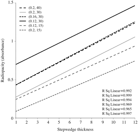
Fig. 1 Linear models with corresponding R2 obtained from various distance/exposure time setups for aluminum step wedge.
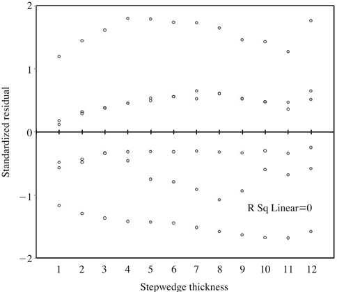
Fig. 2 Magnitude of residue for corresponding thickness of aluminum stepwedge.
Cited by 2 articles
-
Radiopacity of restorative composites by conventional radiograph and digital images with different resolutions
Raquel Venâncio Fernandes Dantas, Hugo Ramalho Sarmento, Rosângela Marques Duarte, Sônia Saeger Meireles Monte Raso, Ana Karina Maciel de Andrade, Maria Luiza Dos Anjos-Pontual
Imaging Sci Dent. 2013;43(3):145-151. doi: 10.5624/isd.2013.43.3.145.Radiopacity of contemporary luting cements using conventional and digital radiography
Seo-Young An, Chang-Hyeon An, Karp-Sik Choi, Kyung-Hoe Huh, Won-Jin Yi, Min-Suk Heo, Sam-Sun Lee, Soon-Chul Choi
Imaging Sci Dent. 2018;48(2):97-101. doi: 10.5624/isd.2018.48.2.97.
Reference
-
1. International Organization for Standardization. ISO 4049: 2009. Dentistry-Polymer based restorative materials. 2009. 4th ed. Geneva: ISO.2. Sur J, Endo A, Matsuda Y, Itoh K, Katoh T, Araki K, et al. A measure for quantifying the radiopacity of restorative resins. Oral Radiol. 2011. 27:22–27.
Article3. Wenzel A, Hintze H, Hørsted-Bindslev P. Discrimination between restorative dental materials by their radiopacity measured in film radiographs and digital images. J Forensic Odontostomatol. 1998. 16:8–13.4. Pedrosa RF, Brasileiro IV, dos Anjos Pontual ML, dos Anjos Pontual A, da Silveira MM. Influence of materials radiopacity in the radiographic diagnosis of secondary caries: evaluation in film and two digital systems. Dentomaxillofac Radiol. 2011. 40:344–350.
Article5. Martinez-Rus F, Garcia AM, de Aza AH, Pradies G. Radiopacity of zirconia-based all-ceramic crown systems. Int J Prosthodont. 2011. 24:144–146.6. Wadhwani C, Hess T, Faber T, Piñeyro A, Chen CS. A descriptive study of the radiographic density of implant restorative cements. J Prosthet Dent. 2010. 103:295–302.
Article7. Ergücü Z, Türkün LS, Onem E, Güneri P. Comparative radiopacity of six flowable resin composites. Oper Dent. 2010. 35:436–440.
Article8. Taşdemir T, Yesilyurt C, Yildirim T, Er K. Evaluation of the radiopacity of new root canal paste/sealers by digital radiography. J Endod. 2008. 34:1388–1390.
Article9. Bodrumlu E, Gungor K. Radiopacity of an endodontic core material. Am J Dent. 2009. 22:157–159.10. Rasimick BJ, Gu S, Deutsch AS, Musikant BL. Measuring the radiopacity of luting cements, dowels, and core build-up materials with a digital radiography system using a CCD sensor. J Prosthodont. 2007. 16:357–364.
Article11. Gu S, Rasimick BJ, Deutsch AS, Musikant BL. Radiopacity of dental materials using a digital X-ray system. Dent Mater. 2006. 22:765–770.
Article12. Tanomaru-Filho M, da Silva GF, Duarte MA, Gonçalves M, Tanomaru JM. Radiopacity evaluation of root-end filling materials by digitization of images. J Appl Oral Sci. 2008. 16:376–379.
Article13. Rasimick BJ, Shah RP, Musikant BL, Deutsch AS. Radiopacity of endodontic materials on film and a digital sensor. J Endod. 2007. 33:1098–1101.
Article14. Baksi BG, Ermis RB. Comparison of conventional and digital radiography for radiometric differentiation of dental cements. Quintessence Int. 2007. 38:e532–e536.15. Catelan A, Padilha AC, Salzedas LM, Coclete GA, dos Santos PH. Effect of radiotherapy on the radiopacity and flexural strength of a composite resin. Acta Odontol Latinoam. 2008. 21:159–162.16. Poorsattar Bejeh Mir A, Poorsattar Bejeh Mir M. How does duration of curing affect radiopacity of dental materials? Imaging Sci Dent. 2012. 42:89–93.17. Watts DC, McCabe JF. Aluminium radiopacity standards for dentistry: an international survey. J Dent. 1999. 27:73–78.
Article18. Nomoto R, Mishima A, Kobayashi K, McCabe JF, Darvell BW, Watts DC, et al. Quantitative determination of radioopacity: equivalence of digital and film X-ray systems. Dent Mater. 2008. 24:141–147.
Article19. Vivan RR, Ordinola-Zapata R, Bramante CM, Bernardineli N, Garcia RB, Hungaro Duarte MA, et al. Evaluation of the radiopacity of some commercial and experimental root-end filling materials. Oral Surg Oral Med Oral Pathol Oral Radiol Endod. 2009. 108:e35–e38.
Article20. White SC, Pharoah MJ. Oral radiology: principles and interpretation. 2009. 6th ed. St. Louis: Mosby-Elsevier;84.
- Full Text Links
-
- Actions
-
Cited
- CITED
-
- Close
- Share
-
- Similar articles
-
- How does duration of curing affect the radiopacity of dental materials?
- Evaluation of radiopacity and discriminability of various fiber reinforced composite posts
- Radiopacity of restorative composites by conventional radiograph and digital images with different resolutions
- A comparative study on radiopacity of root canal sealers
- A comparative study on radiopacity of canal filling and retrograde root-end filling materials
