Brachial Plexopathy due to Myeloid Sarcoma in a Patient With Acute Myeloid Leukemia After Allogenic Peripheral Blood Stem Cell Transplantation
- Affiliations
-
- 1Department of Physical and Rehabilitation Medicine, Samsung Medical Center, Sungkyunkwan University School of Medicine, Seoul, Korea. yays.sung@samsung.com
- KMID: 2165783
- DOI: http://doi.org/10.5535/arm.2013.37.2.280
Abstract
- Myeloid sarcoma is a solid, extramedullary tumor comprising of immature myeloid cells. It may occur in any organ; however, the invasion of peripheral nervous system is rare. Herein, we report the case of myeloid sarcoma on the brachial plexus. A 37-year-old woman with acute myelogenous leukemia achieved complete remission after chemotherapy. One year later, she presented right shoulder pain, progressive weakness in the right upper extremity and hypesthesia. Based on magnetic resonance images (MRI) and electrophysiologic study, a provisional diagnosis of brachial plexus neuritis was done and hence steroid pulse therapy was carried out. Three months later the patient presented epigastric pain. After upper gastrointestinal endoscopy, myeloid sarcoma of gastrointestinal tract was confirmed pathologically. Moreover, 18-fluoride fluorodeoxyglucose positron emission tomography showed a fusiform shaped mass lesion at the brachial plexus overlapping with previous high signal lesion on the MRI. Therefore, we concluded the final diagnosis as brachial plexopathy due to myeloid sarcoma.
MeSH Terms
-
Brachial Plexus
Brachial Plexus Neuritis
Brachial Plexus Neuropathies
Endoscopy, Gastrointestinal
Female
Gastrointestinal Tract
Humans
Hypesthesia
Leukemia, Myeloid, Acute
Magnetic Resonance Spectroscopy
Myeloid Cells
Peripheral Blood Stem Cell Transplantation
Peripheral Nervous System
Positron-Emission Tomography
Sarcoma, Myeloid
Shoulder Pain
Upper Extremity
Figure
-
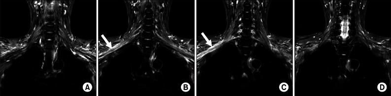
Fig. 1 Magnetic resonance images (coronal, short tau inversion recovery image) from ventral (A) towards dorsal (D) surfaces of the brachial plexus shows diffuse high signal intensity and mild swelling of the right brachial plexus (arrow) from trunk level to cord level. No definite mass lesion was noted within or outside the brachial plexus.

Fig. 2 18-Fluoride fluorodeoxyglucose positron emission tomography (PET) findings. Three-dimensional projection image (A, B) PET/computed tomography fusion axial image (C) reveal hot uptake in stomach, transverse colon, right perihepatic space, right cardiophrenic angle, anterior mediastinum and right brachial plexus. Fusiform shaped mass lesions can be seen at the cord level of right brachial plexus (black and white arrow).
Reference
-
1. Campidelli C, Agostinelli C, Stitson R, Pileri SA. Myeloid sarcoma: extramedullary manifestation of myeloid disorders. Am J Clin Pathol. 2009; 132:426–437. PMID: 19687319.2. Byrd JC, Edenfield WJ, Shields DJ, Dawson NA. Extramedullary myeloid cell tumors in acute nonlymphocytic leukemia: a clinical review. J Clin Oncol. 1995; 13:1800–1816. PMID: 7602369.
Article3. Paydas S, Zorludemir S, Ergin M. Granulocytic sarcoma: 32 cases and review of the literature. Leuk Lymphoma. 2006; 47:2527–2541. PMID: 17169797.
Article4. Bakst R, Jakubowski A, Yahalom J. Recurrent neurotropic chloroma: report of a case and review of the literature. Adv Hematol. 2011; 2011:85240.
Article5. Karam C, Khorsandi A, MacGowan DJ. Clinical reasoning: a 23-year-old woman with paresthesias and weakness. Neurology. 2009; 72:e5–e10. PMID: 19139360.
Article6. Mauermann ML, Angius D, Spinner RJ, Letendre LJ, Amrami KK, Dyck PJ. Isolated granulocytic sarcoma presenting as a brachial plexopathy. J Peripher Nerv Syst. 2008; 13:153–156. PMID: 18601661.7. Byrd JC, Weiss RB. Recurrent granulocytic sarcoma: an unusual variation of acute myelogenous leukemia associated with 8;21 chromosomal translocation and blast expression of the neural cell adhesion molecule. Cancer. 1994; 73:2107–2112. PMID: 7512442.
Article8. Lekos A, Katirji MB, Cohen ML, Weisman R Jr, Harik SI. Mononeuritis multiplex: a harbinger of acute leukemia in relapse. Arch Neurol. 1994; 51:618–622. PMID: 8198473.
- Full Text Links
-
- Actions
-
Cited
- CITED
-
- Close
- Share
-
- Similar articles
-
- Intraparenchymal Myeloid Sarcoma and Subsequent Spinal Myeloid Sarcoma for Acute Myeloblastic Leukemia
- Endobronchial Relapse of Acute Myeloid Leukemia after Allogeneic Stem Cell Transplantation
- The Case Report of a Child with High-Risk Acute Lymphoblastic Leukemia, Treated with Allogenic Peripheral Blood Stem Cell Transplantation
- Four cases of chloroma treated with hematopoietic stem cell transplantation
- Myeloid Sarcoma of Peritoneum in Acute Myeloid Leukemia Patient with Inversion of Chromosome 16
