The Age-Related Orientational Changes of Human Semicircular Canals
- Affiliations
-
- 1Department of Otolaryngology, Eye and ENT Hospital, Fudan University, Shanghai, China.
- 2Research Center, Eye and ENT Hospital, Fudan University, Shanghai, China. peters818@aliyun.com
- 3Key Laboratory of Hearing Medicine, National Ministry of Public Health, Shanghai, China.
- KMID: 2165052
- DOI: http://doi.org/10.21053/ceo.2014.02012
Abstract
OBJECTIVES
Some changes are found in the labyrinth anatomy during postnatal development. Although the spatial orientation of semicircular canals was thought to be stable after birth, we investigated the age-related orientational changes of human semicircular canals during development.
METHODS
We retrospectively studied the computed tomography (CT) images of both ears of 76 subjects ranged from 1 to 70 years old. They were divided into 4 groups: group A (1-6 years), group B (7-12 years), group C (13-18 years), and group D (>18 years). The anatomical landmarks of the inner ear structures were determined from CT images. Their coordinates were imported into MATLAB software for calculating the semicircular canals orientation, angles between semicircular canal planes and the jugular bulb (JB) position. Differences between age groups were analyzed using multivariate statistics. Relationships between variables were analyzed using Pearson analysis.
RESULTS
The angle between the anterior semicircular canal plane and the coronal plane, and the angle between the horizontal semicircular canal plane and the coronal plane were smaller in group D than those in group A (P<0.05). The JB position, especially the anteroposterior position of right JB, correlated to the semicircular canals orientation (P<0.05). However, no statistically significant differences in the angles between ipsilateral canal planes among different age groups were found.
CONCLUSION
The semicircular canals had tendencies to tilt anteriorly simultaneously as a whole with age. The JB position correlated to the spatial arrangement of semicircular canals, especially the right JB. Our calculation method helps detect developmental and pathological changes in vestibular anatomy.
MeSH Terms
Figure
-
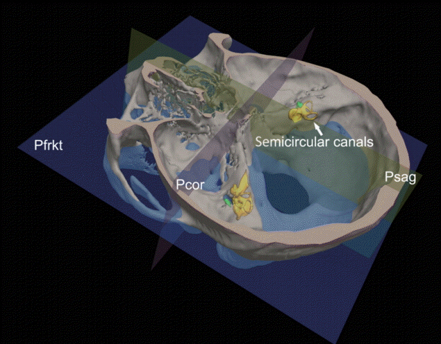
Fig. 1. Standard 3-dimensional coordinate system relative to cranial base we developed. The semicircular canals (arrow) deeply located in the petrous bones. Pfrkt, Frankfurt horizontal plane; Psag, median sagittal plane; Pcor, coronal plane.
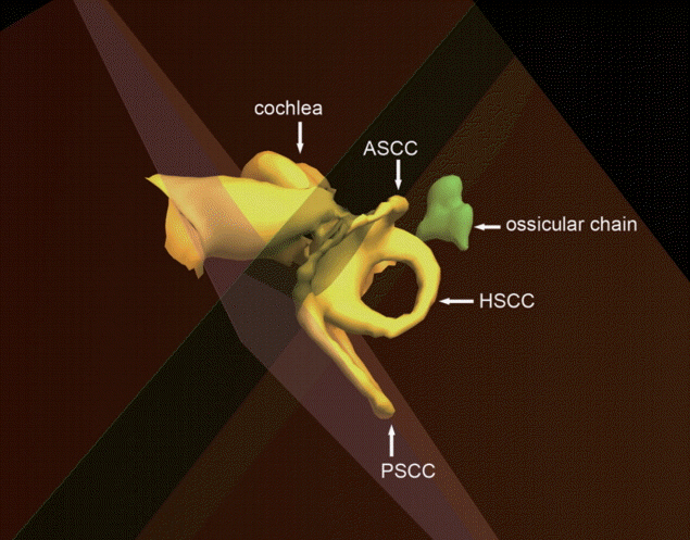
Fig. 2. Right inner ear and ossicular chain. The semicircular canal planes were represented by the planes passing through each canal. ASCC, anterior semicircular canal; PSCC, posterior semicircular canal; HSCC, horizontal semicircular canal.
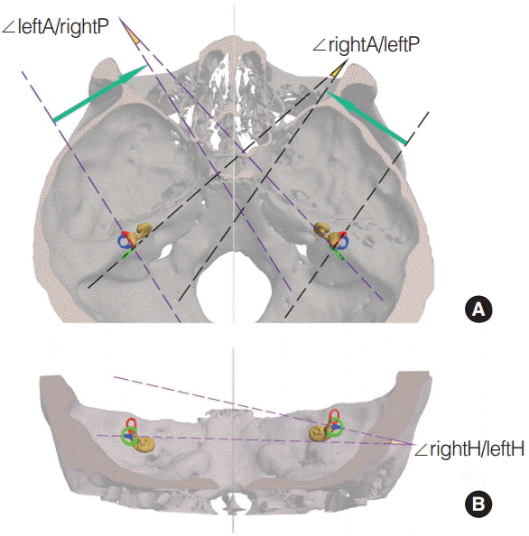
Fig. 3. The angles between contralateral synergistic canals. Panel A is apical view. Panel B is posterior view. The anterior, posterior, and horizontal semicircular canals are red, green and blue colored, respectively. ∠ indicate the angle between 2 planes; A, P, and H, indicate the anterior, posterior, and horizontal semicircular canal plane, respectively.
Reference
-
1. Bradshaw AP, Curthoys IS, Todd MJ, Magnussen JS, Taubman DS, Aw ST, et al. A mathematical model of human semicircular canal geometry: a new basis for interpreting vestibular physiology. J Assoc Res Otolaryngol. 2010; Jun. 11(2):145–59.
Article2. Blanks RH, Curthoys IS, Markham CH. Planar relationships of the semicircular canals in man. Acta Otolaryngol. 1975; Sep-Oct. 80(3-4):185–96.
Article3. Ifediba MA, Rajguru SM, Hullar TE, Rabbitt RD. The role of 3-canal biomechanics in angular motion transduction by the human vestibular labyrinth. Ann Biomed Eng. 2007; Jul. 35(7):1247–63.
Article4. Yang A, Hullar TE. Relationship of semicircular canal size to vestibular-nerve afferent sensitivity in mammals. J Neurophysiol. 2007; Dec. 98(6):3197–205.
Article5. Jeffery N, Spoor F. Prenatal growth and development of the modern human labyrinth. J Anat. 2004; Feb. 204(2):71–92.
Article6. McRackan TR, Reda FA, Rivas A, Noble JH, Dietrich MS, Dawant BM, et al. Comparison of cochlear implant relevant anatomy in children versus adults. Otol Neurotol. 2012; Apr. 33(3):328–34.
Article7. Scott JH. The cranial base. Am J Phys Anthropol. 1958; Sep. 16(3):319–48.
Article8. Barbeito-Andres J, Ventrice F, Anzelmo M, Pucciarelli HM, Sardi ML. Developmental covariation of human vault and base throughout postnatal ontogeny. Ann Anat. 2015; Jan. 197:59–66.9. Lee ST, Mori Y, Minami K, An CH, Park JW, Kwon TG. Does skeletal surgery for asymmetric mandibular prognathism influence the soft tissue contour and thickness? J Oral Maxillofac Surg. 2013; Sep. 71(9):1577–87.
Article10. El Khoury M, Braga J, Dumoncel J, Nancy J, Esclassan R, Vaysse F. The human semicircular canals orientation is more similar to the bonobos than to the Chimpanzees. PLoS One. 2014; Apr. 9(4):e93824.
Article11. Aoki S, Takei Y, Suzuki K, Masukawa A, Arai Y. Planer orientation of the bilateral semicircular canals in dizzy patients. Auris Nasus Larynx. 2012; Oct. 39(5):451–4.
Article12. Kopelman M, Glik A, Greenberg S, Shelef I. Intracranial nonjugular venous pathways: a possible compensatory drainage mechanism. AJNR Am J Neuroradiol. 2013; Jul. 34(7):1348–52.
Article13. Sato H, Sando I, Takahashi H, Fujita S. Torsion of the human semicircular canals and its influence on their angular relationships. Acta Otolaryngol. 1993; Mar. 113(2):171–5.
Article14. Gunz P, Ramsier M, Kuhrig M, Hublin JJ, Spoor F. The mammalian bony labyrinth reconsidered, introducing a comprehensive geometric morphometric approach. J Anat. 2012; Jun. 220(6):529–43.
Article15. Mejdoubi M, Dedouit F, Mokrane FZ, Telmon N. Semicircular canal angulation during fetal life: a computed tomography study of 54 human fetuses. Otol Neurotol. 2015; Apr. 36(4):701–4.16. Suzuki K, Masukawa A, Aoki S, Arai Y, Ueno E. A new coordinates system for cranial organs using magnetic resonance imaging. Acta Otolaryngol. 2010; May. 130(5):568–75.
Article17. Berlin JC, Kirk EC, Rowe TB. Functional implications of ubiquitous semicircular canal non-orthogonality in mammals. PLoS One. 2013; Nov. 8(11):e79585.
Article18. Friedmann DR, Eubig J, McGill M, Babb JS, Pramanik BK, Lalwani AK. Development of the jugular bulb: a radiologic study. Otol Neurotol. 2011; Oct. 32(8):1389–95.19. Dai PD, Zhang HQ, Wang ZM, Sha Y, Wang KQ, Zhang TY. Morphological and positional relationships between the sigmoid sinus and the jugular bulb. Surg Radiol Anat. 2007; Dec. 29(8):643–51.
Article20. Woo CK, Wie CE, Park SH, Kong SK, Lee IW, Goh EK. Radiologic analysis of high jugular bulb by computed tomography. Otol Neurotol. 2012; Sep. 33(7):1283–7.
Article21. Shi L, Wang D, Chu WC, Burwell GR, Wong TT, Heng PA, et al. Automatic MRI segmentation and morphoanatomy analysis of the vestibular system in adolescent idiopathic scoliosis. Neuroimage. 2011; Jan. 54 Suppl 1:S180–8.
Article
- Full Text Links
-
- Actions
-
Cited
- CITED
-
- Close
- Share
-
- Similar articles
-
- Preservation of the Semicircular Canals and Postoperative Hearing in Acoustic Neurinoma Surgery
- A Case of Multiple Bony Dehiscences of Semicircular Canals and Vestibule Presenting as Dizziness and Hearing Loss
- A Case of Benign Paroxysmal Positional Vertigo of the Horizontal Semicircular Canal Following Mastoidectomy
- Mechanism of Downbeat Nystagmus While Normal Subjects HaveHead Upside-down Position in Darkness
- A Case of Lateral Semicircular Canal Dysplasia with Normal Cochlea
