Port Site Metastasis of Breast Cancer after Video-Assisted Thoracic Surgery for Pulmonary Metastasis of Breast Cancer: A Case Report
- Affiliations
-
- 1Department of Radiology, Kangnam Sacred Heart Hospital, Hallym University College of Medicine, Seoul, Korea. imp9653@naver.com
- KMID: 2164830
- DOI: http://doi.org/10.3348/jksr.2016.74.5.331
Abstract
- We reported a case of port site metastasis in a 57-year-old patient who underwent video-assisted thoracic surgery (VATS) resection of pulmonary metastasis from breast cancer. Port site metastasis after VATS is very rare in patients with breast cancer. However, when suspicious lesions are detected near the port site in patients who have undergone VATS for pulmonary metastasis, port site metastasis should be considered in the differential diagnosis.
MeSH Terms
Figure
-
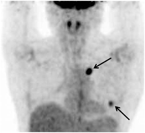
Fig. 1 Positron emission tomography-computed tomography of a 59-year-old woman 2 years after mastectomy. Two pulmonary nodules are seen in the left lung with maximum standardized uptake value of 5.0, suggestive of metastasis (arrows). These nodules were confirmed as metastatic invasive ductal carcinoma from the breast after video-assisted thoracic surgery resection.
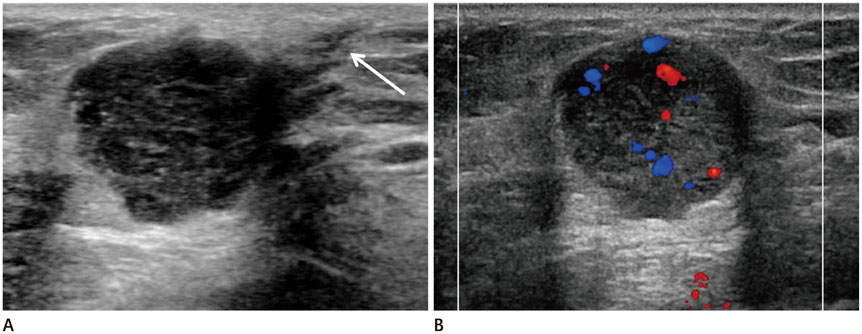
Fig. 2 Ultrasonography (US) performed 6 months after video-assisted thoracic surgery (VATS) resection for evaluation of a palpable mass in the left breast. A. Transverse US scan reveals a 2.0 × 1.8 × 1.9 cm size, circumscribed round mass in lower outer quadrant of left breast around the port insertion area. The mass shows complex cystic and solid echogenicity. Port site scar after VATS (arrow) is demonstrated as subtle linear hypoechoic tract between the mass and the skin. B. On color Doppler US, the mass shows internal vascularity.
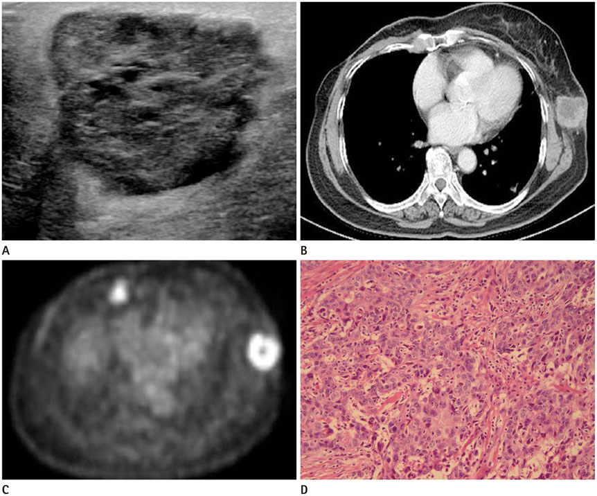
Fig. 3 Follow-up ultrasonography (US), computed tomography (CT), and positron emission tomography-computed tomography (PET-CT) after video-assisted thoracic surgery resection of pulmonary metastasis of breast cancer, and histopathologic image of surgically resected specimen. A. The mass in the lower outer quadrant periphery of the left breast shows an increase in size, as compared with previous US performed 6 months earlier, measuring 3.8 × 3 × 3.4 cm. B. Contrast-enhanced CT demonstrates rim enhancing lobulated mass with central necrosis at the port site. C. PET-CT shows fluorodeoxyglucose (FDG) uptake in the periphery of the mass with maximum standardized uptake value of 5.0. An enlarged right internal mammary lymph node is also noted with increased FDG uptake. D. Photomicrograph shows prominent nuclear pleomorphism, no identifiable tubule formation, and rampant mitotic activity, indicating invasive ductal carcinoma with nuclear grade 3 and modified Bloom-Richardson histologic grade III/III (hematoxylin and eosin stain, × 200).
Reference
-
1. Downey RJ, McCormack P, LoCicero J 3rd. Dissemination of malignant tumors after video-assisted thoracic surgery: a report of twenty-one cases. The Video-Assisted Thoracic Surgery Study Group. J Thorac Cardiovasc Surg. 1996; 111:954–960.2. Thurer RL. Video-assisted thoracic surgery. Ann Thorac Surg. 1993; 56:199–200.3. Walsh GL, Nesbitt JC. Tumor implants after thoracoscopic resection of a metastatic sarcoma. Ann Thorac Surg. 1995; 59:215–216.4. Fry WA, Siddiqui A, Pensler JM, Mostafavi H. Thoracoscopic implantation of cancer with a fatal outcome. Ann Thorac Surg. 1995; 59:42–45.5. Imperatori A, Rotolo N, Gatti M, Nardecchia E, De Monte L, Conti V, et al. Peri-operative complications of video-assisted thoracoscopic surgery (VATS). Int J Surg. 2008; 6:Suppl 1. S78–S81.6. Johnstone PA, Rohde DC, Swartz SE, Fetter JE, Wexner SD. Port site recurrences after laparoscopic and thoracoscopic procedures in malignancy. J Clin Oncol. 1996; 14:1950–1956.7. Jobsen JJ, van der, Ong F, Meerwaldt JH. Synchronous, bilateral breast cancer: prognostic value and incidence. Breast. 2003; 12:83–88.8. Gao X, Fisher SG, Emami B. Risk of second primary cancer in the contralateral breast in women treated for early-stage breast cancer: a population-based study. Int J Radiat Oncol Biol Phys. 2003; 56:1038–1045.9. Hankey BF, Curtis RE, Naughton MD, Boice JD Jr, Flannery JT. A retrospective cohort analysis of second breast cancer risk for primary breast cancer patients with an assessment of the effect of radiation therapy. J Natl Cancer Inst. 1983; 70:797–804.
- Full Text Links
-
- Actions
-
Cited
- CITED
-
- Close
- Share
-
- Similar articles
-
- Thymic Metastasis in Breast Cancer: A Case Report
- A case of stomach metastasis from breast cancer
- Breast Cancer Cutaneous Metastasis at Core Needle Biopsy Site
- Breast Cancer Metastatic to Gluteus Maximus: A Case Report
- An Unusual Case of Gastric Cancer Presenting with Breast Metastasis with Pleomorphic Microcalcifications
