Thymic Metastasis in Breast Cancer: A Case Report
- Affiliations
-
- 1Department of Radiology and the Research Institute of Radiology, University of Ulsan College of Medicine, Asan Medical Center, Seoul, Korea. hhkim@amc.seoul.kr
- 2Department of Pathology, University of Ulsan College of Medicine, Asan Medical Center, Seoul, Korea.
- KMID: 1110736
- DOI: http://doi.org/10.3348/kjr.2007.8.4.360
Abstract
- A malignant tumor is generally believed to be very unlikely to metastasize to the thymus. Only three cases of thymic metastases have been reported so far in the medical literature. We report here a rare case of metastatic breast cancer to the thymus, which was detected by CT and PET scanning, and the metastasis was also confirmed by video-assisted thoracic surgery biopsy. Recognition of an unusual breast cancer metastasis, such as to the thymus, as well as the usual patterns of breast cancer metastasis will facilitate an accurate, prompt diagnosis and its appropriate treatment.
Keyword
MeSH Terms
Figure
-
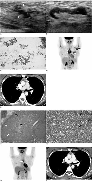
Fig. 1 A 26-year-old woman with thymic metastasis from breast cancer. A. Breast ultrasonography shows a 2.0 × 1.0 × 4.0 cm sized, irregularly shaped, poorly defined hypoechoic mass (arrows) in the 12 o'clock position of the left breast. B. Multiple metastatic lymph nodes in the left level I axilla are shown. A left supraclavicular metastatic lymph node is also noticeable (not shown). C. The specimen obtained on fine-needle aspiration biopsy of the breast mass shows ductal carcinoma (Papanicolau stain, × 400). D. The whole body PET scan shows a focal hypermetabolic lesion (white arrow) in the left upper breast and multiple metastatic lymph nodes (black arrow). In addition, a diffuse hypermetabolic lesion (small arrows) is noticeable in the anterior mediastinum. E. Contrast-enhanced chest CT shows a 1.0 cm sized bulging, nodular soft tissue lesion (arrows) in the anterior mediastinum, which was presumed to be a thymic lesion. F, G. The biopsy specimen, obtained by video-assisted thoracic surgery, shows ductal carcinoma cells (white arrows) and thymic cells (black arrows) in the same fields (Hematoxylin & Eosin staining, × 100). Compared with the specimen obtained by the fine-needle aspiration, the interwoven ductal carcinoma cells (white arrows) with the background thymic cells (black arrows) are basically the same pathology (Hematoxylin & Eosin staining, × 400). The thymic metastasis from primary breast cancer was confirmed. H, I. After two-cycles of chemotherapy, the PET (H) and the CT scans (I) showed that the primary breast mass, the metastatic thymic lesion (arrow) and the metastatic lymphadenopathies were considerably regressed with a partial response.
Cited by 1 articles
-
Gastric Adenocarcinoma with Thymic Metastasis after Curative Resection: A Case Report
Tomoyuki Matsunaga, Hiroaki Saito, Kozo Miyatani, Seigo Takaya, Yoji Fukumoto, Tomohiro Osaki, Masahide Ikeguchi
J Gastric Cancer. 2014;14(3):207-210. doi: 10.5230/jgc.2014.14.3.207.
Reference
-
1. Hayashi S, Hamanaka Y, Sueda T, Yonehara S, Matsuura Y. Thymic metastasis from prostatic carcinoma: report of a case. Surg Today. 1993. 23:632–634.2. Clark SL. The reticulum of lymph nodes in mice studied with the electron microscope. Am J Anat. 1962. 110:217–257.3. Phillips CJ. Case report: metastatic malignant testicular teratoma of the thymus. Br J Radiol. 1994. 67:203–204.4. Nam MS, Chu YC, Choe WS, Kim SJ, Hong SB, Kim YJ, et al. Metastatic follicular thyroid carcinoma to the thymus in a 35-year-old woman. Yonsei Med J. 2002. 43:665–669.5. Middleton G. Involvement of the thymus by metastatic neoplasms. Br J Cancer. 1966. 20:41–46.6. Patanaphan V, Salazar OM, Risco R. Breast cancer: metastatic patterns and their prognosis. South Med J. 1988. 81:1109–1112.7. Ampil FL, Jawahar A, Zamani R, Krishnamsetty RM. Breast cancer metastasis to the medulla oblongata: a case report. Eur J Gynaecol Oncol. 2004. 25:737–738.
- Full Text Links
-
- Actions
-
Cited
- CITED
-
- Close
- Share
-
- Similar articles
-
- Postoperative Radiotherapy in Thymic Carcinoma : A case report
- Sparganosis of the Breast that Mimicked Metastasis: A Case Report
- A case of stomach metastasis from breast cancer
- A Case of Alopecia Neoplastica Metastasis from Breast Carcinoma
- Metastasis of Breast Carcinoma to Intercostal Muscle Detected by Breast MRI: A Case Report
