Ductal Carcinoma In Situ within a Fibroadenoma: Microcalcifications Identified on Mammography Play a Crucial Role in Diagnosis
- Affiliations
-
- 1Department of Diagnostic Radiology, NHIS Ilsan Hospital, Goyang, Korea. toqurquf@empal.com
- 2Department of Pathology, NHIS Ilsan Hospital, Goyang, Korea.
- 3Department of Diagnostic Radiology, Yonsei University College of Medicine, Seoul, Korea.
- KMID: 2164812
- DOI: http://doi.org/10.3348/jksr.2016.74.6.361
Abstract
- Fibroadenoma is a common, benign tumor of the breast, which is rarely associated with an increased risk of carcinoma. We report a case of ductal carcinoma in situ within a fibroadenoma in a 38-year-old woman. The lesion was a 1 cm, circumscribed, ovoid mass with internal calcifications evident on mammography and ultrasound, which is commonly found in fibroadenoma, but the calcifications were fine and linear, which is uncommon. This type of calcification is classified as suspicious by the American College of Radiology Breast Imaging-Reporting And Data System, and it is often correlated with comedo necrosis of ductal carcinoma, and, so, requires immediate pathologic confirmation. In our case, careful analysis of the unusual calcifications led to appropriate intervention and diagnosis. Radiologists should be aware that fibroadenomas can be malignant, and they should look for suspicious microcalcifications within a fibroadenoma.
MeSH Terms
Figure
-

Fig. 1 Mammography, magnification view (A: mediolateral oblique view, B: craniocaudal view). A 1-cm, circumscribed, isodense mass with internal fine, linear microcalcifications is evident in the upper medial quadrant of the right breast (arrow).
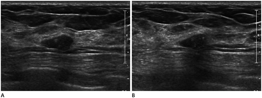
Fig. 2 Ultrasound findings. Ultrasound (A: transverse view, B: longitudinal view) shows a circumscribed ovoid mass with internal echogenic dots in the right breast, 6 cm from the nipple.
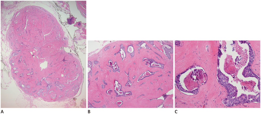
Fig. 3 Microscopic findings of ductal carcinoma in situ within a fibroadenoma. A. Histologic examination shows a well-demarcated fibroadenoma with ductal carcinoma in situ in the lower half (H&E stain, × 12.5). B, C. Comedo-type ductal carcinoma in situ with central necrosis and calcification in a background of hyalinized fibroadenoma. H&E stain, × 40 (B), H&E stain, × 200 (C). H&E = hematoxylin and eosin
Reference
-
1. Deschênes L, Jacob S, Fabia J, Christen A. Beware of breast fibroadenomas in middle-aged women. Can J Surg. 1985; 28:372–374.2. Buzanowski-Konakry K, Harrison EG Jr, Payne WS. Lobular carcinoma arising in fibroadenoma of the breast. Cancer. 1975; 35:450–456.3. Diaz NM, Palmer JO, McDivitt RW. Carcinoma arising within fibroadenomas of the breast. A clinicopathologic study of 105 patients. Am J Clin Pathol. 1991; 95:614–662.4. Baker KS, Monsees BS, Diaz NM, Destouet JM, McDivitt RW. Carcinoma within fibroadenomas: mammographic features. Radiology. 1990; 176:371–374.5. Borecky N, Rickard M. Preoperative diagnosis of carcinoma within fibroadenoma on screening mammograms. J Med Imaging Radiat Oncol. 2008; 52:64–67.6. Kato F, Omatsu T, Matsumura W, Takahashi M, Hosoda M, Takahashi H, et al. Dynamic MR findings of ductal carcinoma in situ within a fibroadenoma. Magn Reson Med Sci. 2011; 10:129–132.7. Shin JH, Choi HY, Lee SN, Kim YJ. Microinvasive ductal carcinoma arising within a fibroadenoma: a case report. Acta Radiol. 2006; 47:643–645.8. Evans A, Pinder S, Wilson R, Sibbering M, Poller D, Elston C, et al. Ductal carcinoma in situ of the breast: correlation between mammographic and pathologic findings. AJR Am J Roentgenol. 1994; 162:1307–1311.9. Stomper PC, Connolly JL. Ductal carcinoma in situ of the breast: correlation between mammographic calcification and tumor subtype. AJR Am J Roentgenol. 1992; 159:483–485.10. American College of Radiology. 2013 ACR BI-RADS Atlas: breast imaging reporting and data system. Reston, VA: American College of Radiology;2014. p. 37–78.
- Full Text Links
-
- Actions
-
Cited
- CITED
-
- Close
- Share
-
- Similar articles
-
- Ductal Carcinoma In Situ in a Fibroadenoma
- Ductal Carcinoma In Situ Detected by Shear Wave Elastography within a Fibroadenoma
- An Unusual Presentation of Extensive Ductal Carcinoma in Situ Accompanying Invasive Ductal Carcinoma on MRI: A Case Report
- Ductal Carcinoma in Situ: US Finding and Its Usefulness
- US-Guided Preoperative Hook-Wire Localization of Nonpalpable Breast Lesions
