Ductal Carcinoma In Situ Detected by Shear Wave Elastography within a Fibroadenoma
- Affiliations
-
- 1Department of Radiology, Istanbul University Cerrahpasa Medical Faculty, Istanbul, Turkey. ustabasioglu@hotmail.com
- 2Department of General Surgery, Istanbul University Cerrahpasa Medical Faculty, Istanbul, Turkey.
- 3Department of Pathology, Istanbul University Cerrahpasa Medical Faculty, Istanbul, Turkey.
Abstract
- Fibroadenoma is the most common breast tumor in women. Malignant transformation occurs rarely within fibroadenoma at older ages. Clinicians, radiologists, and pathologists need to be aware of malignant transformation within fibroadenomas. Radiologic studies play an important role in the diagnosis of fibroadenoma; however, radiologic findings are often nonspecific for malignancy and may appear completely benign. We detected an occult ductal carcinoma in situ that originated inside a fibroadenoma by using shear wave elastography. We report shear wave elastography findings of ductal carcinoma in situ within fibroadenoma and discuss the diagnostic role of this modality.
MeSH Terms
Figure
-
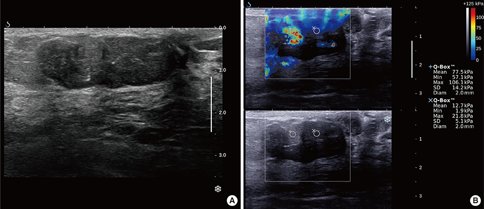
Figure 1 Ductal carcinoma in situ within a fibroadenoma in a 30-year-old female. (A) In the gray scale ultrasonography image, there is a bilobulated lesion parallel to the skin surface. Similar heterogenously hypoechoic appearence is seen in the both compound. A few hyperechoic microcalcification are seen also. (B) In the elastography image, 2 mm diameter region of interest calculates maximum elasticity value of lateral portion of mass as 106.1 kPa, on the other hand it was measured 21.8 kPa on medial portion. Note that display was saturated no shear wave elastography artefact was present.
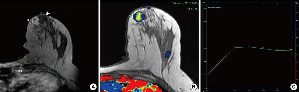
Figure 2 Contrast-enhanced magnetic resonance imaging (MRI) of the breast. (A) Breast MRI examination findings of the lesion. Contrast enhanced T1W oblique reformat image shows significantly enhanced lateral portion (arrow) despite the medial portion (arrowhead) of the lesion. (B) Color coding axial image depicts the lesion clearly. (C) Dynamic evaluation is revealed as early intermediate contrast enhancement followed by late phase washout kinetics (type III) in the lateral portion while slow and minimal persistant enhancement kinetics (type I) in the medial portion (not shown).
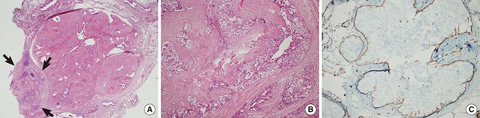
Figure 3 Microscopic findings of ductal carcinoma in situ (DCIS) within fibroadenoma. (A) Magnification view shows DCIS and mild epithelial hyperplasia (arrows) in the fibroadenoma which corresponds to the shear wave elastography and magnetic resonance imaging findings (not all parts of the DCIS portion could be shown due to different slice course) (H&E stain, ×20). (B) Pleomorfic malignant cells are seen within an eosinophilic boundary of basement membran (H&E stain, ×200). (C) This demonstrates the myoepithelial layer around the well defined island of malignant cells with smooth-muscle actin (SMA) immunohistochemistry (×200).
Reference
-
1. Greenberg R, Skornick Y, Kaplan O. Management of breast fibroadenomas. J Gen Intern Med. 1998; 13:640–645.
Article2. Deschênes L, Jacob S, Fabia J, Christen A. Beware of breast fibroadenomas in middle-aged women. Can J Surg. 1985; 28:372–374.3. Ozzello L, Gump FE. The management of patients with carcinomas in fibroadenomatous tumors of the breast. Surg Gynecol Obstet. 1985; 160:99–104.4. Diaz NM, Palmer JO, McDivitt RW. Carcinoma arising within fibroadenomas of the breast: a clinicopathologic study of 105 patients. Am J Clin Pathol. 1991; 95:614–622.
Article5. Abu-Rahmeh Z, Nseir W, Naroditzky I. Invasive ductal carcinoma within fibroadenoma and lung metastases. Int J Gen Med. 2012; 5:19–21.
Article6. Abe H, Hanasawa K, Naitoh H, Endo Y, Tani T, Kushima R. Invasive ductal carcinoma within a fibroadenoma of the breast. Int J Clin Oncol. 2004; 9:334–338.
Article7. Kuijper A, Preisler-Adams SS, Rahusen FD, Gille JJ, van der Wall E, van Diest PJ. Multiple fibroadenomas harbouring carcinoma in situ in a woman with a family history of breast/ovarian cancer. J Clin Pathol. 2002; 55:795–797.
Article8. Giuseppetti GM, Baldassarre S, Marconi E. Color Doppler sonography. Eur J Radiol. 1998; 27:Suppl 2. S254–S258.
Article9. Yang WT, Tse GM. Sonographic, mammographic, and histopathologic correlation of symptomatic ductal carcinoma in situ. AJR Am J Roentgenol. 2004; 182:101–110.
Article10. del Cura JL, Elizagaray E, Zabala R, Legórburu A, Grande D. The use of unenhanced Doppler sonography in the evaluation of solid breast lesions. AJR Am J Roentgenol. 2005; 184:1788–1794.
Article11. Weidner N, Semple JP, Welch WR, Folkman J. Tumor angiogenesis and metastasis: correlation in invasive breast carcinoma. N Engl J Med. 1991; 324:1–8.
Article12. Tiu CM, Chou YH, Chiou SY, Hsu CY, Chen SP, Chiang HR, et al. Development of a carcinoma in situ in a fibroadenoma: color Doppler sonographic demonstration. J Ultrasound Med. 2006; 25:1335–1338.13. Athanasiou A, Tardivon A, Tanter M, Sigal-Zafrani B, Bercoff J, Deffieux T, et al. Breast lesions: quantitative elastography with supersonic shear imaging: preliminary results. Radiology. 2010; 256:297–303.
Article14. Chang JM, Moon WK, Cho N, Yi A, Koo HR, Han W, et al. Clinical application of shear wave elastography (SWE) in the diagnosis of benign and malignant breast diseases. Breast Cancer Res Treat. 2011; 129:89–97.
Article15. Evans A, Whelehan P, Thomson K, McLean D, Brauer K, Purdie C, et al. Quantitative shear wave ultrasound elastography: initial experience in solid breast masses. Breast Cancer Res. 2010; 12:R104.
Article
- Full Text Links
-
- Actions
-
Cited
- CITED
-
- Close
- Share
-
- Similar articles
-
- Diagnostic Performance of Quantitative Shear Wave Ultrasound Elastography for Thyroid Cancer
- Ductal Carcinoma In Situ within a Fibroadenoma: Microcalcifications Identified on Mammography Play a Crucial Role in Diagnosis
- Future of breast elastography
- Non-Invasive Liver Fibrosis Test Using Shear Wave Elastography
- Ultrasound Elastography for Liver Disease with Focus on Hepatic Fibrosis
