Soft Tissue Roasi-Dorfman Disease with Features of IgG4-Related Disease in a Patient with a History of Acute Myeloid Leukemia
- Affiliations
-
- 1Department of Pathology, Yonsei University College of Medicine, Seoul, Korea. nicekyumi@yuhs.ac
- KMID: 2164604
- DOI: http://doi.org/10.4132/jptm.2015.10.08
Abstract
- No abstract available.
MeSH Terms
Figure
-
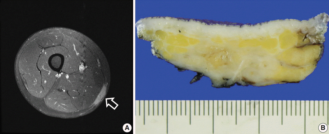
Fig. 1. Imaging and macroscopic findings of soft tissue Rosai-Dorfman disease with features of IgG4-related disease. (A) Magnetic resonance imaging shows a 3.8×2.8 cm enhanced soft tissue mass (arrow) in the subcutaneous layer on the posteromedial side of the right mid-thigh, suggestive of leukemic infiltration. (B) In the excised specimen, a 3.0-cm irregular tan-white mass-like lesion is observed in the subcutis.
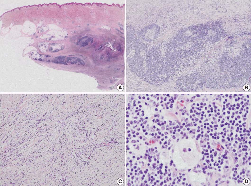
Fig. 2. Microscopic findings of soft tissue Rosai-Dorfman disease with features of IgG4-related disease. (A) In the scanning view, the infiltrative lesion involving the deep dermis and subcutaneous layer is noted. (B) In the low-power view, dense lymphoplasmacytic infiltration with scattered histiocytes is noted. (C) Diffuse lymphoplasmacytic infiltration is noted in the sclerotic stroma. (D) Dilated sinuses are filled with histiocytes containing intact lymphocytes (emperipolesis).

Fig. 3. Immunohistochemical staining results of soft tissue Rosai-Dorfman disease with features of IgG4-related disease. (A) Immunohistochemical staining for CD68 demonstrates histiocytes. (B) Immunohistochemical staining for S100 protein demonstrates emperipolesis. (C) Plasma cell IgG immunoreactivity. (D) Frequently identified IgG4-positive cells.
Reference
-
1. Rosai J, Dorfman RF. Sinus histiocytosis with massive lymphadenopathy: a newly recognized benign clinicopathological entity. Arch Pathol. 1969; 87:63–70.2. Zhang X, Hyjek E, Vardiman J. A subset of Rosai-Dorfman disease exhibits features of IgG4-related disease. Am J Clin Pathol. 2013; 139:622–32.
Article3. Bi Y, Huo Z, Meng Y, et al. Extranodal Rosai-Dorfman disease involving the right atrium in a 60-year-old male. Diagn Pathol. 2014; 9:115.
Article4. Al-Daraji W, Anandan A, Klassen-Fischer M, Auerbach A, Marwaha JS, Fanburg-Smith JC. Soft tissue Rosai-Dorfman disease: 29 new lesions in 18 patients, with detection of polyomavirus antigen in 3 abdominal cases. Ann Diagn Pathol. 2010; 14:309–16.
Article5. Montgomery EA, Meis JM, Frizzera G. Rosai-Dorfman disease of soft tissue. Am J Surg Pathol. 1992; 16:122–9.
Article6. Kong YY, Kong JC, Shi DR, et al. Cutaneous rosai-dorfman disease: a clinical and histopathologic study of 25 cases in China. Am J Surg Pathol. 2007; 31:341–50.7. Young PM, Kransdorf MJ, Temple HT, Mousavi F, Robinson PG. Rosai-Dorfman disease presenting as multiple soft tissue masses. Skeletal Radiol. 2005; 34:665–9.
Article8. Kuo TT, Chen TC, Lee LY, Lu PH. IgG4-positive plasma cells in cutaneous Rosai-Dorfman disease: an additional immunohistochemical feature and possible relationship to IgG4-related sclerosing disease. J Cutan Pathol. 2009; 36:1069–73.
Article9. Deshpande V, Zen Y, Chan JK, et al. Consensus statement on the pathology of IgG4-related disease. Mod Pathol. 2012; 25:1181–92.10. Thomas CG, Patel RM, Bergfeld WF. Cytophagic and S-100 protein immunoreactive myeloid leukemia cutis. J Cutan Pathol. 2010; 37:390–5.
Article
- Full Text Links
-
- Actions
-
Cited
- CITED
-
- Close
- Share
-
- Similar articles
-
- Unusual Manifestation of Immunoglobulin G4-Related Disease Involving the Upper Arm: A Case Report
- Rosai-Dorfman Disease: Report of a Case Associated with IgG4-Related Sclerotic Lesions
- Myeloid Sarcoma of Peritoneum in Acute Myeloid Leukemia Patient with Inversion of Chromosome 16
- A t(3;3)(q21;q26) Acute Myeloid Leukemia with the Philadelphia Chromosome as a Secondary Change
- Rosai-Dorfman Disease in the Breast with Increased IgG4 Expressing Plasma Cells: A Case Report
