Bone Transport for Reconstruction in Benign Bone Tumors
- Affiliations
-
- 1Department of Orthopaedic Surgery, Chonnam National University Hospital, Chonnam National University Medical School, Gwangju, Korea. stjung@jnu.ac.kr
- 2Department of Orthopaedic Surgery, Yonsei University College of Medicine, Seoul, Korea.
- KMID: 2164552
- DOI: http://doi.org/10.4055/cios.2015.7.2.248
Abstract
- BACKGROUND
The aim of this study was to assess the results of using the Ilizarov apparatus to transport bones in the treatment of benign bone tumors.
METHODS
Seven patients (six males and one female) with benign bone tumors were treated by bone transport with an Ilizarov apparatus at our institution. Their mean age at surgery was 14.4 years (range, 4.8 to 36.9 years). The histological diagnoses were osteofibrous dysplasia (4), giant-cell tumor (1), intraosseous cavernous hemangioma (1), and aneurysmal bone cyst (1). Three radiological indices were used for evaluating the results: an external fixation index, a distraction index, and a maturation index. The bone and functional results were evaluated according to the Association for the Study and Application of the Method of Ilizarov classification.
RESULTS
Five patients had bone union at the reconstructed site, one patient had a local recurrence, and the other had a nonunion at the docking site. The mean length of distraction was 7.3 cm (range, 5.1 to 12.1 cm). The mean external fixation index was 26.0 day/cm (range, 19.8 to 32.5 day/cm), the distraction index was 9.6 day/cm (range, 6.8 to 12.0 day/cm), and the maturation index was 14.9 day/cm (range, 8.0 to 22.5 day/cm). Ultimately, the bone and the functional results were rated excellent in six cases and good in one case.
CONCLUSIONS
Bone transport using the Ilizarov apparatus is a good treatment option in patients with bone defects after the resection of an active or aggressive benign bone tumor.
Keyword
MeSH Terms
Figure
-
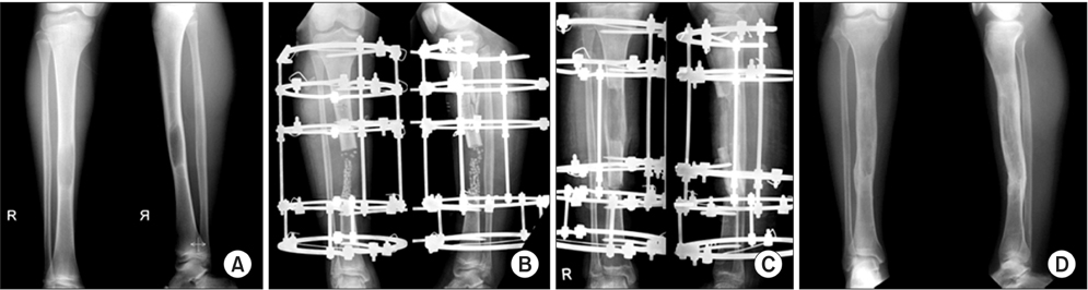
Fig. 1 (A) A 14-year-old male with an osteolytic lesion of the tibia shaft. (B) We resected the lesion with calcium sulfate grafting and applied the Ilizarov apparatus. The histological diagnosis was cavernous hemangioma of the bone. (C) Distraction was done. (D) Union of the transported bone was achieved.

Fig. 2 (A) A 36-year-old male patient with an osteolytic lesion of the distal femur. (B) Tumor was resected and proximal femur was docked to the distal femur. The histological diagnosis was giant cell tumor. (C) Lengthening was done at proximal from the docking site. (D) Docking site and distracted bone union was achieved.
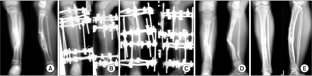
Fig. 3 (A) A 4-year-old male patient with an osteolytic lesion of the tibia shaft. (B) Tumor was resected with tricorticotomy. The histological diagnosis was osteofibrous dysplasia. (C) Bone transport was done at proximal and distal corticotomy site. (D) Bone union and angular deformity. For protection of stress fracture, we applied long leg splint. (E) The last follow-up radiograph shows the remodeled tibia without recurrence.
Reference
-
1. Ilizarov GA. The tension-stress effect on the genesis and growth of tissues. Part I. the influence of stability of fixation and soft-tissue preservation. Clin Orthop Relat Res. 1989; (238):249–281.2. Ilizarov GA. The tension-stress effect on the genesis and growth of tissues: Part II. The influence of the rate and frequency of distraction. Clin Orthop Relat Res. 1989; (239):263–285.3. Ilizarov GA, Green SA. The transosseous osteosynthesis: theoretical and clinical aspects of the regeneration and growth of tissue. Berlin: Springer-Verlag;1992.4. Song HR, Cho SH, Koo KH, Jeong ST, Park YJ, Ko JH. Tibial bone defects treated by internal bone transport using the Ilizarov method. Int Orthop. 1998; 22(5):293–297.5. Robert Rozbruch S, Weitzman AM, Tracey Watson J, Freudigman P, Katz HV, Ilizarov S. Simultaneous treatment of tibial bone and soft-tissue defects with the Ilizarov method. J Orthop Trauma. 2006; 20(3):197–205.6. Shalaby S, Shalaby H, Bassiony A. Limb salvage for osteosarcoma of the distal tibia with resection arthrodesis, autogenous fibular graft and Ilizarov external fixator. J Bone Joint Surg Br. 2006; 88(12):1642–1646.7. Watanabe K, Tsuchiya H, Yamamoto N, et al. Over 10-year follow-up of functional outcome in patients with bone tumors reconstructed using distraction osteogenesis. J Orthop Sci. 2013; 18(1):101–109.8. Tsuchiya H, Tomita K, Minematsu K, Mori Y, Asada N, Kitano S. Limb salvage using distraction osteogenesis: a classification of the technique. J Bone Joint Surg Br. 1997; 79(3):403–411.9. Paley D, Catagni MA, Argnani F, Villa A, Benedetti GB, Cattaneo R. Ilizarov treatment of tibial nonunions with bone loss. Clin Orthop Relat Res. 1989; (241):146–165.10. Kapukaya A, Subasi M, Kandiya E, Ozates M, Yilmaz F. Limb reconstruction with the callus distraction method after bone tumor resection. Arch Orthop Trauma Surg. 2000; 120(3-4):215–218.11. Nishida J, Shimamura T. Methods of reconstruction for bone defect after tumor excision: a review of alternatives. Med Sci Monit. 2008; 14(8):RA107–RA113.12. Laffosse JM, Accadbled F, Abid A, Kany J, Darodes P, Sales De. Reconstruction of long bone defects with a vascularized fibular graft after tumor resection in children and adolescents: thirteen cases with 50-month follow-up. Rev Chir Orthop Reparatrice Appar Mot. 2007; 93(6):555–563.13. Shin KH, Park HJ, Yoo JH, Hahn SB. Reconstructive surgery in primary malignant and aggressive benign bone tumor of the proximal humerus. Yonsei Med J. 2000; 41(3):304–311.14. Tsuchiya H, Morsy AF, Matsubara H, Watanabe K, Abdel-Wanis ME, Tomita K. Treatment of benign bone tumours using external fixation. J Bone Joint Surg Br. 2007; 89(8):1077–1083.15. Karita M, Tsuchiya H, Sakurakichi K, Tomita K. Osteofibrous dysplasia treated with distraction osteogenesis: a report of two cases. J Orthop Sci. 2004; 9(5):516–520.16. El-Alfy B, El-Mowafi H, Kotb S. Bifocal and trifocal bone transport for failed limb reconstruction after tumour resection. Acta Orthop Belg. 2009; 75(3):368–373.17. Green SA, Jackson JM, Wall DM, Marinow H, Ishkanian J. Management of segmental defects by the Ilizarov intercalary bone transport method. Clin Orthop Relat Res. 1992; (280):136–142.18. Oh CW, Shetty GM, Song HR, et al. Submuscular plating after distraction osteogenesis in children. J Pediatr Orthop B. 2008; 17(5):265–269.19. Kocaoglu M, Eralp L, Bilen FE, Balci HI. Fixator-assisted acute femoral deformity correction and consecutive lengthening over an intramedullary nail. J Bone Joint Surg Am. 2009; 91(1):152–159.
