Primary Mucosa-associated Lymphoid Tissue Lymphoma Metachronously Involving Esophagus and Stomach
- Affiliations
-
- 1Department of Internal Medicine, Dongguk University Ilsan Hospital, Goyang, Korea. gangmali@naver.com
- 2Department of Pathology, Dongguk University Ilsan Hospital, Goyang, Korea.
- KMID: 2164333
- DOI: http://doi.org/10.4166/kjg.2016.67.5.257
Abstract
- Mucosa-associated lymphoid tissue (MALT) lymphoma is found in various organs as extranodal B cell lymphoma. The gastrointestinal tract is the most commonly involved extranodal site in MALT lymphoma. However, primary esophageal MALT lymphoma is very rare. In addition, few cases with metachronous gastric involvement have been reported. A 55-year-old man was diagnosed with MALT lymphoma by surveillance esophagogastroduodenoscopy. A 5 cm esophageal submucosal tumor-like lesion was incidentally revealed by screening esophagogastroduodenoscopy two years prior. Esophagogastroduodenoscopy showed a cylindrically elongated submucosal mass with normal overlying mucosa in the mid esophagus. He underwent surgery to confirm the diagnosis. The pathologic diagnosis was esophageal MALT lymphoma. He was treated with radiation, which achieved complete remission. Esophagogastroduodenoscopy and chest computed tomography were performed every three to six months, with no evidence of recurrence for 18 months. After 21 months, several elevated gastric erosions were found on the great curvature and posterior sides of the midbody and confirmed as MALT lymphoma pathologically. Here we report a case with MALT lymphoma metachronously involving the esophagus and stomach.
MeSH Terms
Figure
-
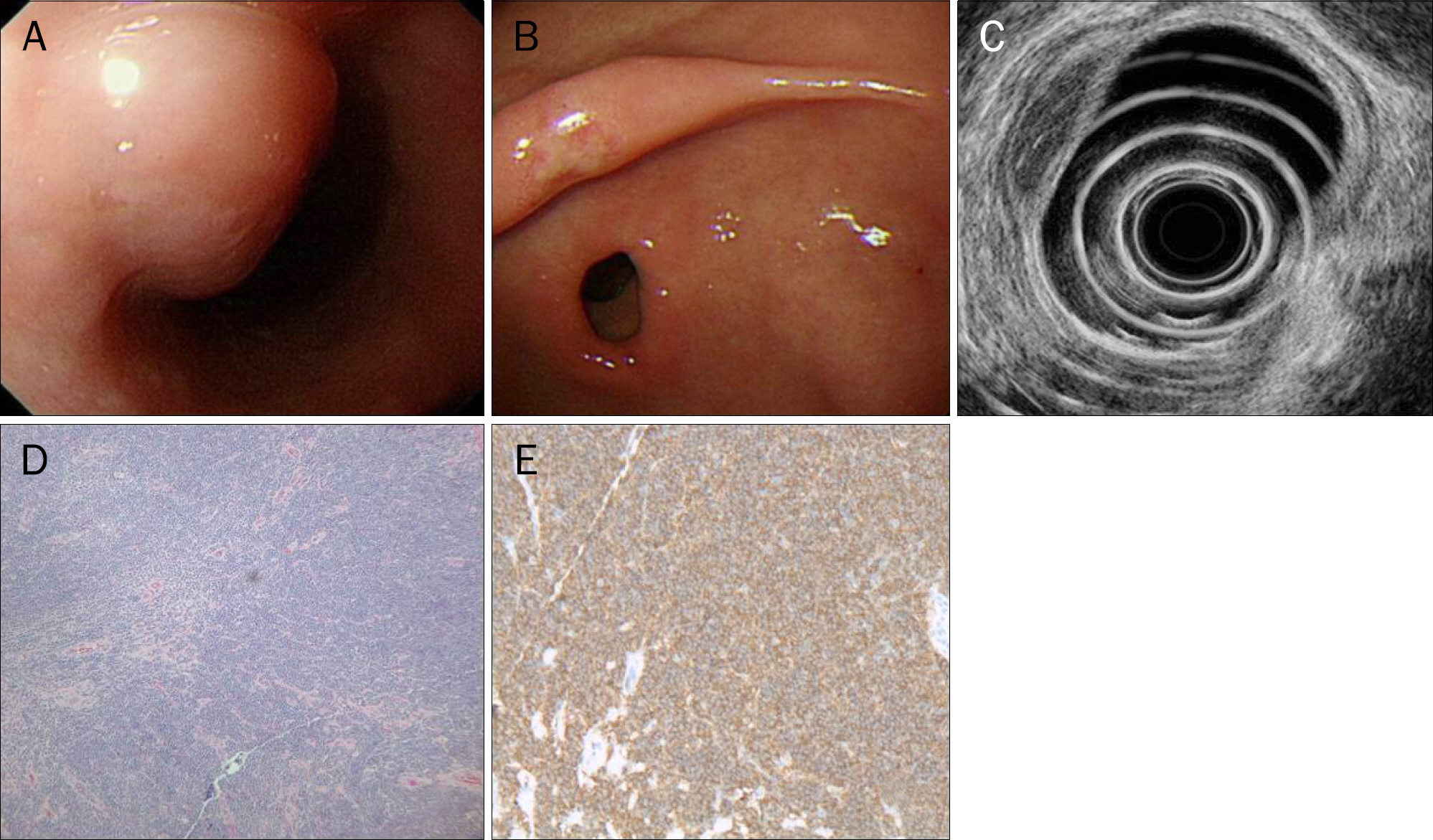
Fig. 1. Intial endoscopic and pathologic findings of the esophageal mass. (A) Esophagogastroduodenoscopy reveals elongated esophageal submucosal tumor-like lesion of the esophagus 25–30 cm from the incisor teeth. (B) Esophagogastroduodenoscopy reveals erosion on the antrum. (C) EUS shows an ovoid homogenous hypoechoic mass located in the submucosal layers. (D) Diffuse infiltration of atypical lymphoid cells (H&E,×100). (E) Positive immunoreactivity for the CD20 protein (immunohistochemistry, ×400).
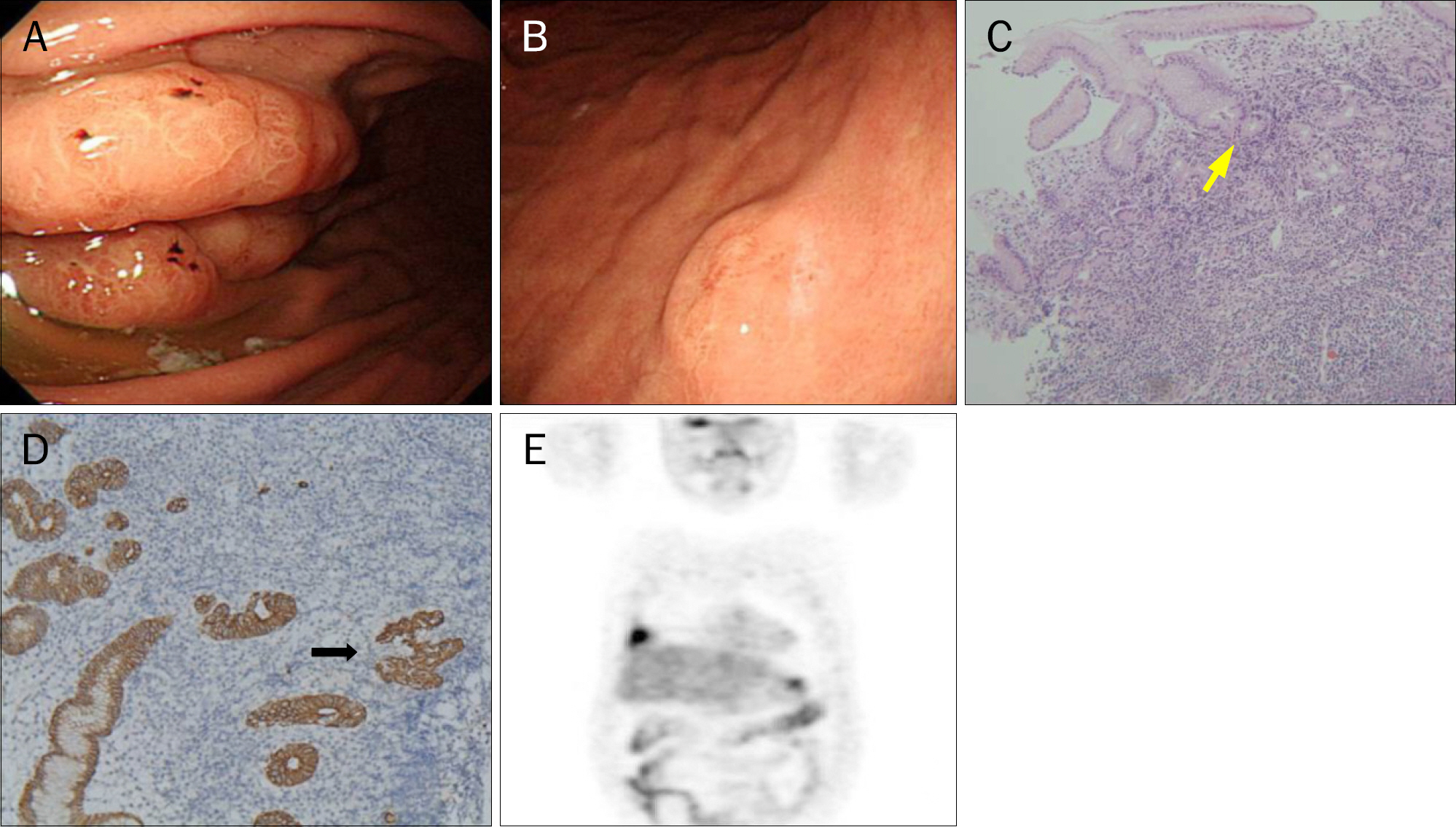
Fig. 2. Follow-up endoscopic, pathologic and PET findings of gastric lesion. (A) Esophagogastroduodenoscopy shows mucosal fold fusion on the greater curvature of the midbody. (B) Esophagogastroduodenoscopy shows erosion on the posterior wall of the midbody. (C) The lymphoid infiltrate in mucosa-associated lymphoid tissue (MALT) lymphoma extends deeper into the lamina propria (H&E, ×200; arrow, lymphoepithelial lesion). (D) Immunohistochemistry for CD20 shows many B-cells and contains lymphoepithelial lesions in which neoplastic B-cells infiltrated (immunohistochemistry, ×400; arrow, lymphoepithelial lesion). (E) Whole body PET-CT shows diffuse hypermetabolic lesion (maximum standardized uptake value=4.1) in the greater curvature side wall of body in stomach and hypermetabolic lesion (maximum standardized uptake value=5.4) in the right middle lung subpleural space.
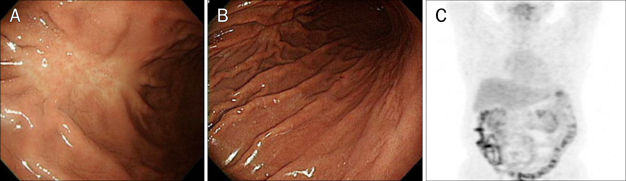
Fig. 3. Esophagogastroduodenoscopic and PET findings following chemotherapy. (A) Esophagogastroduodenoscopy shows whitish mucosal change by scar. (B) Esophagogastroduodenoscopy shows no mucosal change on the posterior wall of the midbody. (C) Abnormal hypermetabolic lesions are not detected in the stomach and lung using PET-CT.
Reference
-
References
1. Hosaka S, Nakamura N, Akamatsu T, et al. A case of primary low grade mucosa associated lymphoid tissue (MALT) lymphoma of the oesophagus. Gut. 2002; 51:281–284.
Article2. Kim GH, Choi BG, Lee JN, et al. 2 cases of gastric mucosa-associated lymphoid tissue lymphoma presenting as a submucosal tumorlike lesion. Korean J Gastroenterol. 2010; 56:103–108.
Article3. Lepicard A, Lamarque D, Lévy M, et al. Duodenal mucosa-associated lymphoid tissue lymphoma: treatment with oral cyclophosphamide. Am J Gastroenterol. 2000; 95:536–539.
Article4. Madabhavi I, Patel A, Revannasiddaiah S, et al. Primary esophageal Burkitt's lymphoma: a rare case report and review of literature. Gastroenterol Hepatol Bed Bench. 2014; 7:230–237.5. Ghai S, Pattison J, Ghai S, O'Malley ME, Khalili K, Stephens M. Primary gastrointestinal lymphoma: spectrum of imaging findings with pathologic correlation. Radiographics. 2007; 27:1371–1388.
Article6. Isaacson P, Wright DH. Malignant lymphoma of mucosa-associated lymphoid tissue. A distinctive type of B-cell lymphoma. Cancer. 1983; 52:1410–1416.
Article7. Amer MH, el-Akkad S. Gastrointestinal lymphoma in adults: clinical features and management of 300 cases. Gastroenterology. 1994; 106:846–858.
Article8. Herrmann R, Panahon AM, Barcos MP, Walsh D, Stutzman L. Gastrointestinal involvement in non-Hodgkin's lymphoma. Cancer. 1980; 46:215–222.
Article9. Schmid C, Vazquez JJ, Diss TC, Isaacson PG. Primary B-cell mucosa-associated lymphoid tissue lymphoma presenting as a solitary colorectal polyp. Histopathology. 1994; 24:357–362.
Article10. Hosaka S, Akamatsu T, Nakamura S, et al. Mucosa-associated lymphoid tissue (MALT) lymphoma of the rectum with chromosomal translocation of the t(11;18)(q21;q21) and an additional aberration of trisomy 3. Am J Gastroenterol. 1999; 94:1951–1954.
Article11. Jung JG, Kang HW, Hahn SJ, Choi JS, Kim EJ. Primary mucosa-associated lymphoid tissue lymphoma of the esophagus, manifesting as a submucosal tumor. Korean J Gastroenterol. 2013; 62:117–121.
Article12. Zucca E, Bertoni F, Roggero E, Cavalli F. The gastric marginal zone B-cell lymphoma of MALT type. Blood. 2000; 96:410–419.
Article13. Weston AP, Cherian R, Horvat RT, Lawrinenko V, Dixon A, McGregor D. Mucosa-associated lymphoid tissue (MALT) in Barrett's esophagus: prospective evaluation and association with gastric MALT, MALT lymphoma, and Helicobacter pylori. Am J Gastroenterol. 1997; 92:800–804.14. Hayashi M, Ueda K, Tanaka T, et al. Mucosa-associated lymphoid tissue (MALT) lymphoma arising in the esophagus, stomach, and lung. Gen Thorac Cardiovasc Surg. 2011; 59:826–830.
Article15. Wotherspoon AC, Doglioni C, Diss TC, et al. Regression of primary low-grade B-cell gastric lymphoma of mucosa-associated lymphoid tissue type after eradication of Helicobacter pylori. Lancet. 1993; 342:575–577.
Article
- Full Text Links
-
- Actions
-
Cited
- CITED
-
- Close
- Share
-
- Similar articles
-
- Mucosa-Associated Lymphoid Tissue Lymphoma of the Esophagus Coexistent with Bronchus-Associated Lymphoid Tissue Lymphoma of the Lung
- A Case of Mucosa Associated Lymphoid Tissue (MALT) Lymphoma Originated from Uterine Endometrium
- A Case of Mucosa-Associated Lymphoid Tissue Lymphoma in the Esophagus Accompanied by Bronchus-Associated Lymphoid Tissue Lymphoma
- A case report of the Pulmonary Malignant Lymphomaof the mucosa-associated lymphoid tissue(MALT)
- Mucosa-associated lymphoid tissue lymphoma on right lower eyelid previously diagnosed as lymphoid hyperplasia
