J Periodontal Implant Sci.
2016 Apr;46(2):128-134. 10.5051/jpis.2016.46.2.128.
Rescue of a periodontally compromised tooth by non-surgical treatment: a case report
- Affiliations
-
- 1Department of Periodontology, Seoul National University School of Dentistry, Seoul, Korea. guy@snu.ac.kr
- KMID: 2161989
- DOI: http://doi.org/10.5051/jpis.2016.46.2.128
Abstract
- PURPOSE
This article describes a case of the successful non-surgical management of a periodontally compromised maxillary premolar.
METHODS
A combination therapy, including root planing, occlusal adjustment, and tooth splinting, was applied. Clinical and radiographic examinations were performed during the 16-month follow-up period.
RESULTS
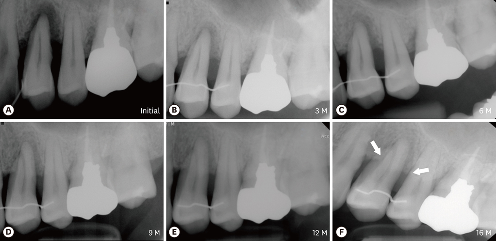
All periodontal parameters were improved. There were dramatic decreases (3-6 mm) in the probing pocket depth, tooth mobility, and marginal bone loss. Interestingly, gradual resolution of the periapical radiolucency and alveolar bone regeneration were observed in the radiographs, and the periodontal condition was maintained during the follow-up period.
CONCLUSIONS
Within the limits of this study, these results demonstrate the importance of natural tooth preservation through proper periodontal treatment and occlusal adjustment of the periodontally compromised tooth, which is typically targeted for tooth extraction and dental implantation.
MeSH Terms
Figure
Reference
-
1. Armitage GC. Development of a classification system for periodontal diseases and conditions. Northwest Dent. 2000; 79:31–35.
Article2. Heitz-Mayfield LJ, Schätzle M, Löe H, Bürgin W, Anerud A, Boysen H, et al. Clinical course of chronic periodontitis. II. Incidence, characteristics and time of occurrence of the initial periodontal lesion. J Clin Periodontol. 2003; 30:902–908.3. Heitz-Mayfield LJ, Trombelli L, Heitz F, Needleman I, Moles D. A systematic review of the effect of surgical debridement vs non-surgical debridement for the treatment of chronic periodontitis. J Clin Periodontol. 2002; 29:Suppl 3. 92–102.
Article4. Wiebe CB, Putnins EE. The periodontal disease classification system of the American Academy of Periodontology--an update. J Can Dent Assoc. 2000; 66:594–597.5. Loesche WJ, Giordano JR, Soehren S, Kaciroti N. The nonsurgical treatment of patients with periodontal disease: results after five years. J Am Dent Assoc. 2002; 133:311–320.6. Sanz-Sánchez I, Ortiz-Vigón A, Matos R, Herrera D, Sanz M. Clinical efficacy of subgingival debridement with adjunctive erbium:yttrium-aluminum-garnet laser treatment in patients with chronic periodontitis: a randomized clinical trial. J Periodontol. 2015; 86:527–535.
Article7. Skurska A, Dolinska E, Pietruska M, Pietruski JK, Dymicka V, Kemona H, et al. Effect of nonsurgical periodontal treatment in conjunction with either systemic administration of amoxicillin and metronidazole or additional photodynamic therapy on the concentration of matrix metalloproteinases 8 and 9 in gingival crevicular fluid in patients with aggressive periodontitis. BMC Oral Health. 2015; 15:63.
Article8. Smiley CJ, Tracy SL, Abt E, Michalowicz BS, John MT, Gunsolley J, et al. Evidence-based clinical practice guideline on the nonsurgical treatment of chronic periodontitis by means of scaling and root planing with or without adjuncts. J Am Dent Assoc. 2015; 146:525–535.
Article9. Smiley CJ, Tracy SL, Abt E, Michalowicz BS, John MT, Gunsolley J, et al. Systematic review and meta-analysis on the nonsurgical treatment of chronic periodontitis by means of scaling and root planing with or without adjuncts. J Am Dent Assoc. 2015; 146:508–24.e5.
Article10. Branschofsky M, Beikler T, Schäfer R, Flemming TF, Lang H. Secondary trauma from occlusion and periodontitis. Quintessence Int. 2011; 42:515–522.11. Davies SJ, Gray RJ, Linden GJ, James JA. Occlusal considerations in periodontics. Br Dent J. 2001; 191:597–604.
Article12. Jin LJ, Cao CF. Clinical diagnosis of trauma from occlusion and its relation with severity of periodontitis. J Clin Periodontol. 1992; 19:92–97.
Article13. Glickman I, Smulow JB. Further observations on the effects of trauma from occlusion in humans. J Periodontol. 1967; 38:280–293.
Article14. Ericsson I, Lindhe J. Effect of longstanding jiggling on experimental marginal periodontitis in the beagle dog. J Clin Periodontol. 1982; 9:497–503.
Article15. Hallmon WW. Occlusal trauma: effect and impact on the periodontium. Ann Periodontol. 1999; 4:102–108.
Article16. Harrel SK, Nunn ME. The effect of occlusal discrepancies on periodontitis. II. Relationship of occlusal treatment to the progression of periodontal disease. J Periodontol. 2001; 72:495–505.
Article17. Gher ME. Changing concepts. The effects of occlusion on periodontitis. Dent Clin North Am. 1998; 42:285–299.18. Lindhe J, Nyman S. The role of occlusion in periodontal disease and the biological rationale for splinting in treatment of periodontitis. Oral Sci Rev. 1977; 10:11–43.19. Harrel SK. Occlusal forces as a risk factor for periodontal disease. Periodontol 2000. 2003; 32:111–117.
Article20. Hallmon WW, Harrel SK. Occlusal analysis, diagnosis and management in the practice of periodontics. Periodontol 2000. 2004; 34:151–164.
Article21. Bourgeois M. Panoramic radiography for the general practitioner. Ont Dent. 1994; 71:29–30.22. Rushton VE, Horner K, Worthington HV. Routine panoramic radiography of new adult patients in general dental practice: relevance of diagnostic yield to treatment and identification of radiographic selection criteria. Oral Surg Oral Med Oral Pathol Oral Radiol Endod. 2002; 93:488–495.
Article
- Full Text Links
- Actions
-
Cited
- CITED
-
- Close
- Share
- Similar articles
-
- Stability of periodontally compromised teeth after splint and non-surgical therapy: two cases followed-up for 1 to 3 years
- Automatic detection of periodontal compromised teeth in digital panoramic radiographs using faster regional convolutional neural networks
- A STUDY ON THE CONFIGURATION OF THE BRISTLE FOR THE PROSTHETIC AND PERIODONTALLY INVOLVED PATIENT.
- Full mouth rehabilitation with a few remaining teeth and implants for a patient with chronic periodontitis: a case report
- Causes of failures of long-term used double crown denture and new rehabilitation with dental implant and tooth combined denture using remaining teeth and implants


