Detection of Perivalvular Abscess with Late Gadolinium-Enhanced MR Imaging in a Patient with Infective Endocarditis
- Affiliations
-
- 1Department of Radiology, Samsung Medical Center, Sungkyunkwan University School of Medicine, Seoul, Korea. yhchoe@skku.edu
- 2HVSI Imaging Center, Heart Vascular Stroke Institute, Samsung Medical Center, Sungkyunkwan University School of Medicine, Seoul, Korea.
- KMID: 2161374
- DOI: http://doi.org/10.13104/imri.2016.20.1.75
Abstract
- We report a case of perivalvular abscess in a 66-year-old man with infective endocarditis, diagnosed by late gadolinium-enhanced (LGE) cardiovascular magnetic resonance (CMR) imaging. No clinical features suspicious of infective endocarditis were noted, however, transthoracic echocardiography revealed non-specific echogenic focal wall thickening at mitral-aortic intervalvular fibrosa. Perivalvular abscess in the aortic valve was demonstrated as focal wall thickening between the anterior mitral leaflet and the non-coronary cusp of the aortic valve with peripheral enhancement and central low signal intensity on LGE CMR imaging. Other features suggestive of infective endocarditis, such as neither vegetation nor valvular perforation were present. The perivalvular abscess did not grow after intensive intravenous antibiotics therapy, and the patient was discharged without surgical treatment. CMR with LGE provided an early accurate diagnosis of perivalvular abscess. The diagnosis of perivalvular abscess using LGE CMR imaging was not previously reported in Korea.
Keyword
MeSH Terms
Figure
-
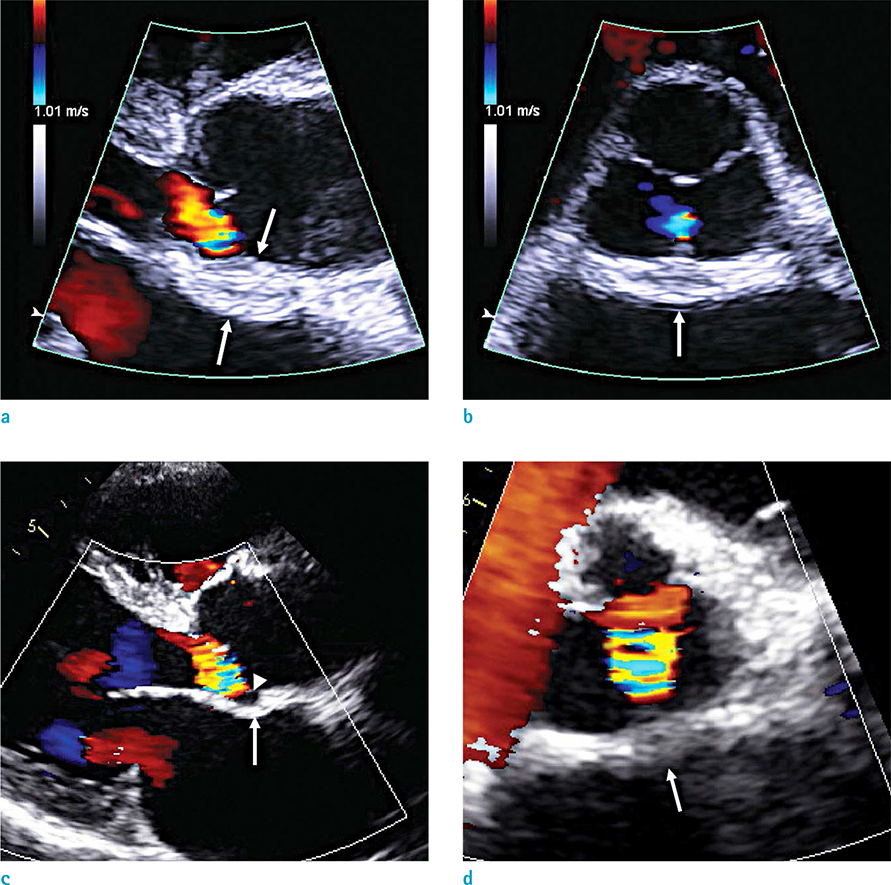
Fig. 1 Transthoracic echocardiography findings in a 66-year-old male. (a, b) Initial echocardiography shows thickening of aortic perivalvular area (arrows). (c, d) Follow-up study 37 days after the first echocardiography shows improved thickness of the lesion (arrows) and an echo-lucent focus (arrowhead).
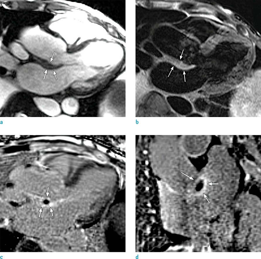
Fig. 2 MR imaging findings of perivalvular abscess in a 66-year-old male, two days after initial echocardiography. (a) 3-chamber view cine MR image shows focal wall thickening between the non-coronary cusp of the aortic valve and the anterior mitral leaflet (arrows). (b) 3-chamber view T2-weighted image shows slightly increased signal intensity of the focal wall thickening (arrows). (c, d) gadolinium contrast-enhanced delayed myocardial images demonstrate peripheral late gadolinium enhancements (arrows) with central hypointensity.
Reference
-
1. Baddour LM, Wilson WR, Bayer AS, et al. Infective endocarditis in adults: diagnosis, antimicrobial therapy, and management of complications: a scientific statement for healthcare professionals from the american heart association. Circulation. 2015; 132:1435–1486.2. Habib G, Badano L, Tribouilloy C, et al. Recommendations for the practice of echocardiography in infective endocarditis. Eur J Echocardiogr. 2010; 11:202–219.3. Bruun NE, Habib G, Thuny F, Sogaard P. Cardiac imaging in infectious endocarditis. Eur Heart J. 2014; 35:624–632.4. Feuchtner GM, Stolzmann P, Dichtl W, et al. Multislice computed tomography in infective endocarditis: comparison with transesophageal echocardiography and intraoperative findings. J Am Coll Cardiol. 2009; 53:436–444.5. Harris KM, Ang E, Lesser JR, Sonnesyn SW. Cardiac magnetic resonance imaging for detection of an abscess associated with prosthetic valve endocarditis: a case report. Heart Surg Forum. 2007; 10:E186–E187.6. Sverdlov AL, Taylor K, Elkington AG, Zeitz CJ, Beltrame JF. Images in cardiovascular medicine. Cardiac magnetic resonance imaging identifies the elusive perivalvular abscess. Circulation. 2008; 118:e1–e3.7. Dursun M, Yilmaz S, Yilmaz E, et al. The utility of cardiac MRI in diagnosis of infective endocarditis: preliminary results. Diagn Interv Radiol. 2015; 21:28–33.8. Saby L, Le Dolley Y, Laas O, et al. Early diagnosis of abscess in aortic bioprosthetic valve by 18F-fluorodeoxyglucose positron emission tomography-computed tomography. Circulation. 2012; 126:e217–e220.9. Pizzi MN, Roque A, Fernandez-Hidalgo N, et al. Improving the diagnosis of infective endocarditis in prosthetic valves and intracardiac devices with 18F-fluordeoxyglucose positron emission tomography/computed tomography angiography: initial results at an infective endocarditis referral center. Circulation. 2015; 132:1113–1126.
- Full Text Links
-
- Actions
-
Cited
- CITED
-
- Close
- Share
-
- Similar articles
-
- Myocardial Infarction Caused by Coronary Artery Compression From Perivalvular Abscess
- Diagnosis of Right Ventricular Vegetation on Late Gadolinium-Enhanced MR Imaging in a Pediatric Patient after Repair of a Ventricular Septal Defect
- Streptococcus Constellatus Community Acquired Pneumonia with Subsequent Isolated Pulmonic Valve Endocarditis and Abscess Formation in a Structurally Normal Heart
- Detectability of Hepatocellular Carcinoma: Comparison of Gd-DT PA-Enhanced and SPIO-Enhanced MR Imaging
- Left Ventricular Pseudoaneurysm after Surgery for Infective Endocarditis with Annular Abscess: A case report
