J Cardiovasc Ultrasound.
2016 Mar;24(1):79-83. 10.4250/jcu.2016.24.1.79.
Stress-Induced Cardiomyopathy Presenting as Shock
- Affiliations
-
- 1Division of Cardiology, Department of Internal Medicine, Kangbuk Samsung Hospital, Sungkyunkwan University School of Medicine, Seoul, Korea. kcmd.sung@samsung.com
- 2Department of Cardiovascular Surgery, Kangbuk Samsung Hospital, Sungkyunkwan University School of Medicine, Seoul, Korea.
- KMID: 2161002
- DOI: http://doi.org/10.4250/jcu.2016.24.1.79
Abstract
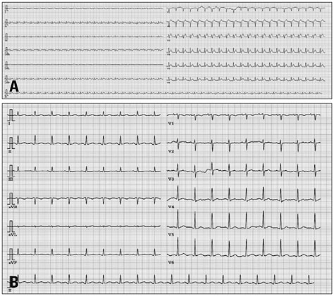
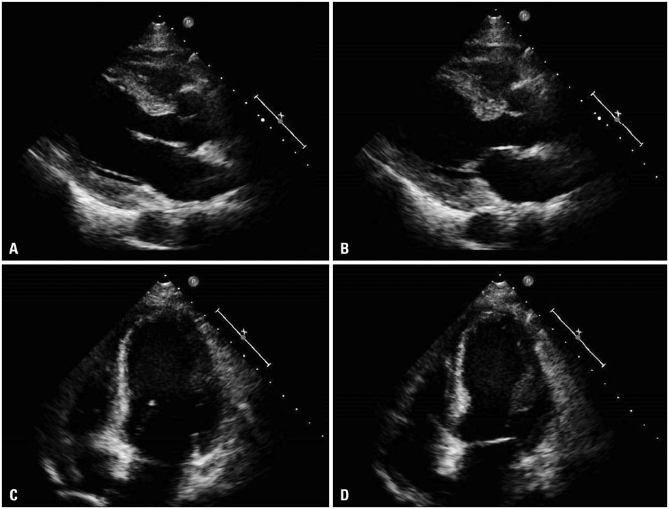
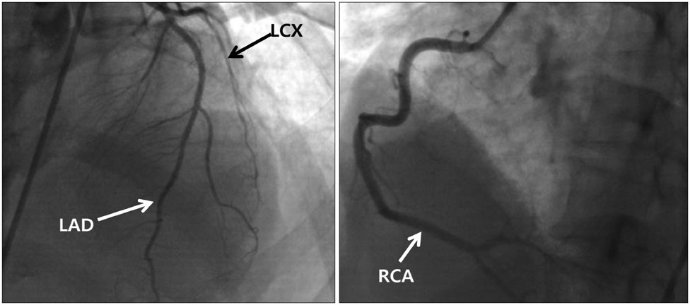
- Stress-induced cardiomyopathy has become a more recognized and reported entity. It can be caused by emotional or physical stress, which causes excessive catecholamine release. Typically, the clinical course is benign with conservative treatment being effective. However, stress-induced cardiomyopathy can be fatal. A 41-year-old female presented with cardiogenic shock followed by sudden back pain. Initial echocardiographic finding showed severely decreased ejection fraction with akinesia at all mid-to-apical walls with relatively preserved basal wall contractility. The coronary artery was intact on coronary angiography. Cardiac resuscitation and extra-corporeal membrane oxygenation was needed to manage the cardiogenic shock. Recovery was complete after 2 weeks.
Keyword
MeSH Terms
Figure
Reference
-
1. Wan SH, Liang JJ. Takotsubo cardiomyopathy: etiology, diagnosis, and optimal management. Res Rep Clin Cardiol. 2014; 5:297–303.2. Cesário V, Loureiro MJ, Pereira H. [Takotsubo cardiomyopathy in a cardiology department]. Rev Port Cardiol. 2012; 31:603–608.3. Bybee KA, Prasad A. Stress-related cardiomyopathy syndromes. Circulation. 2008; 118:397–409.4. Boland TA, Lee VH, Bleck TP. Stress-induced cardiomyopathy. Crit Care Med. 2015; 43:686–693.5. Templin C, Ghadri JR, Diekmann J, Napp LC, Bataiosu DR, Jaguszewski M, Cammann VL, Sarcon A, Geyer V, Neumann CA, Seifert B, Hellermann J, Schwyzer M, Eisenhardt K, Jenewein J, Franke J, Katus HA, Burgdorf C, Schunkert H, Moeller C, Thiele H, Bauersachs J, Tschöpe C, Schultheiss HP, Laney CA, Rajan L, Michels G, Pfister R, Ukena C, Böhm M, Erbel R, Cuneo A, Kuck KH, Jacobshagen C, Hasenfuss G, Karakas M, Koenig W, Rottbauer W, Said SM, Braun-Dullaeus RC, Cuculi F, Banning A, Fischer TA, Vasankari T, Airaksinen KE, Fijalkowski M, Rynkiewicz A, Pawlak M, Opolski G, Dworakowski R, MacCarthy P, Kaiser C, Osswald S, Galiuto L, Crea F, Dichtl W, Franz WM, Empen K, Felix SB, Delmas C, Lairez O, Erne P, Bax JJ, Ford I, Ruschitzka F, Prasad A, Lüscher TF. Clinical features and outcomes of takotsubo (stress) cardiomyopathy. N Engl J Med. 2015; 373:929–938.6. Deshmukh A, Kumar G, Pant S, Rihal C, Murugiah K, Mehta JL. Prevalence of takotsubo cardiomyopathy in the United States. Am Heart J. 2012; 164:66–71.e1.7. Akashi YJ, Goldstein DS, Barbaro G, Ueyama T. Takotsubo cardiomyopathy: a new form of acute, reversible heart failure. Circulation. 2008; 118:2754–2762.8. Bybee KA, Kara T, Prasad A, Lerman A, Barsness GW, Wright RS, Rihal CS. Systematic review: transient left ventricular apical ballooning: a syndrome that mimics ST-segment elevation myocardial infarction. Ann Intern Med. 2004; 141:858–865.9. Sharkey SW, Lesser JR, Zenovich AG, Maron MS, Lindberg J, Longe TF, Maron BJ. Acute and reversible cardiomyopathy provoked by stress in women from the United States. Circulation. 2005; 111:472–479.10. Tsuchihashi K, Ueshima K, Uchida T, Oh-mura N, Kimura K, Owa M, Yoshiyama M, Miyazaki S, Haze K, Ogawa H, Honda T, Hase M, Kai R, Morii I. Angina Pectoris-Myocardial Infarction Investigations in Japan. Transient left ventricular apical ballooning without coronary artery stenosis: a novel heart syndrome mimicking acute myocardial infarction. Angina pectoris-myocardial infarction investigations in Japan. J Am Coll Cardiol. 2001; 38:11–18.11. Prasad A, Lerman A, Rihal CS. Apical ballooning syndrome (tako-tsubo or stress cardiomyopathy): a mimic of acute myocardial infarction. Am Heart J. 2008; 155:408–417.12. Eitel I, von Knobelsdorff-Brenkenhoff F, Bernhardt P, Carbone I, Muellerleile K, Aldrovandi A, Francone M, Desch S, Gutberlet M, Strohm O, Schuler G, Schulz-Menger J, Thiele H, Friedrich MG. Clinical characteristics and cardiovascular magnetic resonance findings in stress (takotsubo) cardiomyopathy. JAMA. 2011; 306:277–286.13. Wagner A, Schulz-Menger J, Dietz R, Friedrich MG. Long-term follow-up of patients paragraph sign with acute myocarditis by magnetic paragraph sign resonance imaging. MAGMA. 2003; 16:17–20.
- Full Text Links
- Actions
-
Cited
- CITED
-
- Close
- Share
- Similar articles
-
- Stress-induced Cardiomyopathy Associated with Non-Small Cell Lung Cancer Presenting as Hyponatremia
- Stress-induced Cardiomyopathy Following Cesarean Delivery with Hemorrhagic Shock: A Case Report
- Stress-Induced Cardiomyopathy: The Role of Echocardiography
- Recurrent fetal postpartum stress induced cardiomyopathy after normal vaginal delivery
- A case of stress-induced cardiomyopathy with an "inverted Takotsubo" contractile pattern


